Educational content only. Not personalised medical advice. Clinical decisions remain the responsibility of the treating clinician.
Visual Pathway Lesions and Field Defects
Visual field defects follow the site of damage along the visual pathway. Chiasmal compression, often by pituitary adenoma, causes visual deficits that may improve after surgery [S5] [S11]. Retrochiasmal lesions cause homonymous field defects, found in about 8% of stroke patients, which often stop people reading, driving or working [S2]. Rehabilitation focuses on compensation, because no widely accepted therapy restores the lost field [S7] [S12].
Key points - Negative visual symptoms such as hemianopia and field defects point to a true stroke rather than a stroke mimic [S8]. - Some homonymous hemianopias improve, usually within days of infarction, but they remain stable after 3 months [S2]. - After endoscopic pituitary surgery, field defects improved in 80.8% but recovered completely in only 40.4% [S5]. - Compensatory rehabilitation gives the most consistent functional benefit; restorative training has not shown a clear field gain in RCTs [S7] [S12]. - Reading training improves reading speed in homonymous field defects (meta-analysis of RCTs) [S9].
Evidence last checked: 2026-10-17 (PubMed abstracts).
Red flags
- Sudden hemianopia, visual loss, gaze palsy or visual inattention: these are more frequent in stroke than in mimics [S8]. Treat as possible acute stroke (see Refer when).
- A field defect with thinning of the nerve fibre and ganglion cell layers on OCT, as seen with para-chiasmal tumours [S11].
- A patient with a homonymous defect who still drives: some are unsafe, and field extent alone does not predict on-road performance [S3].
Recognise it
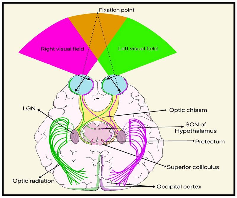
- Patterns differ between conditions (idiopathic intracranial hypertension, optic neuropathy, chiasmal compression and stroke) [S6].
- Retrochiasmal disease (optic tract, lateral geniculate nucleus, optic radiation, occipital lobe) produces homonymous field defects [S2].
- Stroke: in 13 studies (9248 patients), hemianopia, visual field defect, gaze palsy, eye movement disorder and visual inattention were more common in stroke than in mimics, while blurred vision and non-systematised visual trouble were more common in mimics [S8].
- Chiasmal lesions: in a meta-analysis of 97 studies, patients with para-chiasmal tumours had thinner peripapillary nerve fibre layer, macular ganglion cell complex and ganglion cell-inner plexiform layer than controls [S11].
Confirm it
- Perimetry: Humphrey 30-2 and 24-2 programs were most commonly used, followed by Goldmann kinetic perimetry [S6]. There are no guidelines for perimetry in neurological conditions, and central programs may miss early peripheral loss [S6].
- OCT can support diagnosis and prognosis in para-chiasmal lesions; thicker pre-operative nerve fibre layer was associated with good visual recovery (mean difference 11.35 µm) [S11]. MRI remains the primary diagnostic tool for pituitary tumours [S11].
- Neuroimaging and its timing are not covered by these abstracts; follow local stroke and neuro-oncology pathways.
Manage it
Before acting on this section, check the national and local guidelines, product information and regulatory approvals that apply where you practise. See the medical disclaimer.
Tier A (primary care / no specialist)
- Any patient recognised with a field defect after stroke needs prompt referral for assessment and consideration of visual rehabilitation [S1].
- Rehabilitation should match each patient's needs; options include optical therapy, eye movement therapy and visual field restitution [S1].
- Ask about driving: some people with homonymous defects have significant deficits in skills needed for safe driving [S3].
Tier B (general eye clinic)
- Map the field with an appropriate program, considering peripheral testing [S6].
- Use OCT where available in suspected chiasmal disease [S11].
- Counsel that improvement after stroke usually occurs early and that defects are stable after 3 months [S2].
- Offer reading training: a meta-analysis of 9 randomised studies found improved reading speed (SMD 0.30), and reading training itself was effective (SMD 0.35) [S9].
Tier C (specialist centre)
- Visual scanning training: in a pilot RCT (87 patients), visual search training improved vision-related quality of life, field area did not change in any arm, and Fresnel prisms caused adverse events, typically headache, in 69% [S4]. In the larger SEARCH RCT (161 patients, 34 UK stroke units), scanning training did not differ from sham at 26 weeks; both groups improved, possibly through a placebo effect [S10].
- Restorative training: home motion-discrimination training in the blind field (RCT, 48 patients) gave no significant difference from training in the sighted field [S7]. Restorative and neuromodulation approaches give variable results [S12].
- Pituitary adenoma: endoscopic endonasal surgery improved acuity in 67.5% and fields in 80.8%, with deterioration in 4.50% and 2.3% respectively (35 case series) [S5].
- Driving: offer individualised on-road or simulator assessment, because no test yet predicts who is safe [S3].
Refer when
| Situation | Urgency |
|---|---|
| Sudden field loss or other new visual symptoms suggesting stroke | Immediate, same-day stroke pathway assessment [S8] [EIC expert opinion] |
| Field defect suggesting chiasmal compression | Urgent, for MRI; timing per your local service or specialist advice [S11] |
| Stable field defect after stroke | Routine rehabilitation referral; timing per your local service or specialist advice; prompt referral is advised [S1] |
| Patient with a homonymous defect who wishes to drive | Routine, for fitness-to-drive assessment; timing per your local service or specialist advice [S3] |
Pitfalls
- Dismissing "blurred vision" in a stroke patient without testing fields; field defects are typical of stroke [S8].
- Relying only on central perimetry [S6].
- Promising field recovery from training [S7] [S10] [S12].
- Using visual field extent alone to judge driving safety [S3].
Evidence notes
| Source | Date | Design |
|---|---|---|
| [S1] | 2010 | Practical review for general practitioners |
| [S2] | 2011 | Narrative review (textbook chapter) |
| [S3] | 2016 | Narrative literature review |
| [S4] | 2017 | Pilot RCT, 87 patients (UK) |
| [S5] | 2017 | Meta-analysis of 35 case series |
| [S6] | 2018 | Systematic review, 330 studies |
| [S7] | 2021 | RCT, 48 patients |
| [S8] | 2023 | Systematic review of 13 observational studies |
| [S9] | 2024 | Meta-analysis of 9 randomised studies |
| [S10] | 2025 | Double-blind RCT (SEARCH), 161 patients; UK charity funded |
| [S11] | 2025 | Systematic review and meta-analysis, 97 studies |
| [S12] | 2026 | Systematic review, 28 studies of mixed design |
- Conflict: the pilot RCT found better quality of life with scanning training than standard care [S4], but the larger sham-controlled SEARCH trial found no difference [S10]; the pilot compared against information only.
- The specific shapes of chiasmal and retrochiasmal defects (bitemporal, homonymous quadrantanopia, macular sparing) are not described in these abstracts and need a source.
- Not covered by these sources: localisation rules for field defects, imaging protocols, prism fitting details, and low-resource rehabilitation services.
Related articles
References
See the numbered source list below. Each marker [S#] links to its PubMed and DOI record.
Sources
- [S1] Luu S, Lee AW, Daly A, Chen CS Visual field defects after stroke--a practical guide for GPs. Australian family physician. (2010). PubMed 20628665
- [S2] Fraser JA, Newman NJ, Biousse V Disorders of the optic tract, radiation, and occipital lobe. Handbook of clinical neurology. (2011). PubMed 21601068 · doi:10.1016/b978-0-444-52903-9.00014-5
- [S3] Bowers AR Driving with homonymous visual field loss: a review of the literature. Clinical & experimental optometry. (2016). PubMed 27535208 · doi:10.1111/cxo.12425
- [S4] Rowe FJ, Conroy EJ, Bedson E, Cwiklinski E, Drummond A, García-Fiñana M, et al. A pilot randomized controlled trial comparing effectiveness of prism glasses, visual search training and standard care in hemianopia. Acta neurologica Scandinavica. (2017). PubMed 28028819 · doi:10.1111/ane.12725
- [S5] Muskens IS, Zamanipoor Najafabadi AH, Briceno V, Lamba N, Senders JT, van Furth WR, et al. Visual outcomes after endoscopic endonasal pituitary adenoma resection: a systematic review and meta-analysis. Pituitary. (2017). PubMed 28643208 · doi:10.1007/s11102-017-0815-9
- [S6] Hepworth LR, Rowe FJ Programme choice for perimetry in neurological conditions (PoPiN): a systematic review of perimetry options and patterns of visual field loss. BMC ophthalmology. (2018). PubMed 30200926 · doi:10.1186/s12886-018-0912-1
- [S7] Cavanaugh MR, Blanchard LM, McDermott M, Lam BL, Tamhankar M, Feldon SE Efficacy of Visual Retraining in the Hemianopic Field after Stroke: Results of a Randomized Clinical Trial. Ophthalmology. (2021). PubMed 33242498 · doi:10.1016/j.ophtha.2020.11.020
- [S8] Helboe KS, Eddelien HS, Kruuse C Visual symptoms in acute stroke - A systematic review of observational studies. Clinical neurology and neurosurgery. (2023). PubMed 37163931 · doi:10.1016/j.clineuro.2023.107749
- [S9] Maeyama T, Okada H, Sakai S The effects of rehabilitative interventions on reading disorders caused by homonymous visual field defects: a meta-analysis focusing on improvement in reading speed. Acta neurologica Belgica. (2024). PubMed 37572263 · doi:10.1007/s13760-023-02327-6
- [S10] Rowe FJ, Brayshaw E, Brown M, Chatterjee K, Drummond A, Hazelton C, et al. A randomized controlled trial of Scanning Eye trAining as a Rehabilitation Choice for Hemianopia after stroke (SEARCH). International journal of stroke : official journal of the International Stroke Society. (2025). PubMed 40083185 · doi:10.1177/17474930251330140
- [S11] Lim KSA, Tng WXA, Theng WB, Lee BTK, Chin CF, Li KZ, et al. The role of optical coherence tomography in the evaluation of para-chiasmal lesions: a systematic review and meta-analysis. Frontiers in ophthalmology. (2025). PubMed 41669541 · doi:10.3389/fopht.2025.1691582
- [S12] Díaz-Marín N, López-de-la-Fuente C, Marcén Y, Pérez-Rey J, Bilbao C, López-de-la-Fuente MJ Interventions for Visual Field Loss After Acquired Brain Injury: A Systematic Review. Life (Basel, Switzerland). (2026). PubMed 42652963 · doi:10.3390/life16081275