Educational content only. Not personalised medical advice. Clinical decisions remain the responsibility of the treating clinician.
Retinal Vein Occlusion
Retinal vein occlusion (RVO) is the second most common retinal vascular disorder, affecting an estimated 28.06 million people aged 30-89 years worldwide in 2015 [S5]. Vision is lost mainly through macular oedema [S7] [S9], and intravitreal anti-VEGF is the recommended first-line treatment [S6] [S9]. RVO is also a marker of cardiovascular risk, including stroke [S10] [S6].
Key points - Hypertension is the strongest risk factor for RVO (odds ratio 2.82) [S5]; RVO patients have a higher risk of stroke (risk ratio 1.38) [S10]. - Anti-VEGF improves vision in macular oedema from both central (CRVO) and branch (BRVO) occlusion (high- to moderate-certainty evidence) [S1] [S7]. - Ranibizumab and bevacizumab gave comparable short-term results in direct comparisons [S12]. - Best outcomes come when intravitreal treatment starts early [S9]; laser is an option where reliable monitoring is not feasible [S6]. - Routine thrombophilia screening is not recommended [S4].
Evidence last checked: 2026-10-17 (PubMed abstracts).
Red flags
- Extensive non-perfusion (ischaemic CRVO). In a small RAVE trial substudy, non-perfusion was profound at baseline and increased in all 12 eyes despite anti-VEGF, by about 8.1% of the perfused retina per year [S2]. The duration of non-perfusion is a crucial prognostic factor requiring timely intervention [S6].
- Uncontrolled vascular risk. RVO raised the risk of both ischaemic (risk ratio 1.37) and haemorrhagic (risk ratio 1.55) stroke [S10].
- Raised intraocular pressure on treatment. Intraocular pressure elevation was more frequent in the anti-VEGF network meta-analysis (odds ratio 1.924) and warrants monitoring [S11]; steroids increased cataract and raised pressure [S7].
- Delay in starting treatment worsens outcomes [S9].
Recognise it: who is at risk
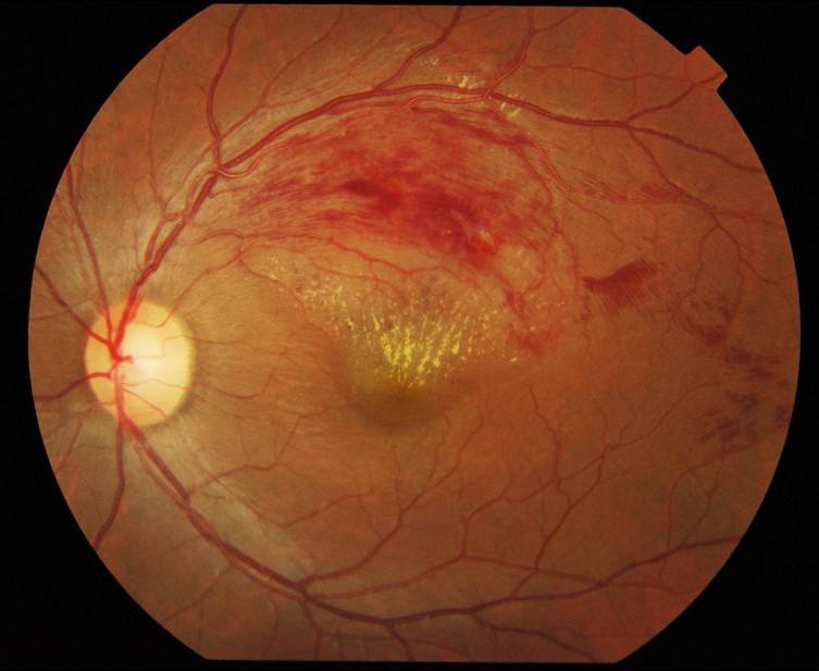
- Global prevalence in people aged 30-89 years was 0.77% for any RVO, 0.64% for BRVO and 0.13% for CRVO; prevalence rises with age and does not differ by sex [S5].
- Hypertension is the strongest risk factor [S5]; cardiovascular disease is common in the elderly with retinal vascular disease [S6].
- Acute leakage produces the hallmark signs of retinal haemorrhage and oedema with marked retinal thickening [S6].
- Persistent macular oedema develops in 60% of eyes with BRVO; untreated, only 14% of eyes with chronic oedema keep 20/40 or better [S3].
Confirm it
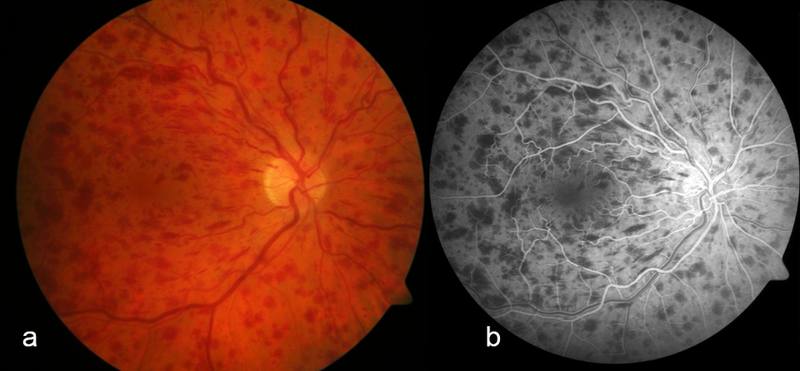
- Trials measured central retinal thickness on OCT to assess macular oedema and its response [S7] [S1].
- Fluorescein angiography, and increasingly OCT angiography, evaluate retinal ischaemia and the risk of late complications [S6]. Wide-field angiography showed the extent of non-perfusion in ischaemic CRVO [S2].
- A basic systemic workup is advised to recognise systemic associations [S6]. Systematic thrombophilia screening cannot be recommended (all evidence low quality) [S4].
Manage it
Before acting on this section, check the national and local guidelines, product information and regulatory approvals that apply where you practise. See the medical disclaimer.
Tier A (primary care / no specialist)
- Check and treat blood pressure and other vascular risk factors; hypertension is the strongest risk factor and stroke risk is raised [S5] [S10].
- Do not order routine thrombophilia tests [S4].
- Refer promptly to an eye clinic: early intravitreal treatment gives the best outcomes [S9].
Tier B (general eye clinic)
- Macular grid laser for BRVO. In one RCT, grid laser vs observation improved the chance of gaining 10 or more letters at 36 months (risk ratio 1.75, moderate-quality evidence) [S3]. Early or subthreshold laser was not supported [S3]. Laser may have an adjunctive role in BRVO but not in CRVO [S9].
- Laser where monitoring is not feasible. EURETINA lists destructive laser as an option if reliable monitoring is not feasible (expert guideline) [S6].
- Bevacizumab. Four RCTs (759 participants) found no significant difference from ranibizumab in visual acuity change at 6 months (mean difference 0.61 letters) [S12]. Doses are not stated in the abstract; follow local formularies.
Tier C (specialist centre)
- Anti-VEGF for CRVO. Compared with sham, anti-VEGF made a gain of 15 or more letters at 6 months 2.71 times more likely and cut the risk of losing 15 or more letters by 80% (high-quality evidence) [S1].
- Anti-VEGF for BRVO. Anti-VEGF outperformed sham, macular laser (mean 9.63 letters better at 6 months) and intravitreal steroid (mean 9.15 letters better at 12 months, high-certainty evidence) [S7]. The earlier grid laser review found insufficient evidence of anti-VEGF benefit over laser [S3]; the newer review is more favourable to anti-VEGF [S7].
- Choice of drug. A 2025 network meta-analysis of 17 RCTs (4,272 eyes) found a pooled gain of 15.14 letters with anti-VEGF and ranked faricimab most effective, with aflibercept, ranibizumab and bevacizumab also beneficial [S11].
- Steroids. Anti-VEGF is recommended over intravitreal corticosteroid because it is more effective and has fewer ocular adverse events [S9]. Steroids may be considered in patients with systemic cardiovascular risk [S6]; they increase cataract and pressure [S7].
- Long-term care. RVO is chronic and monitoring should be individualised [S6]. Across 76 data sets, vision improved by 16.1 letters at 5 years with long-term intravitreal treatment [S8]. Surgery is advisable only for very few patients [S6].
Refer when
| Situation | Urgency |
|---|---|
| Sudden painless vision loss with retinal haemorrhages | Urgent, within a week, to an eye clinic with OCT [EIC expert opinion] |
| RVO with new vessels on the iris, disc or retina, or raised pressure with pain | Same-day to an eye specialist |
| Any RVO: vascular risk assessment | Routine, with primary care for blood pressure and cardiovascular review [S5] [S10]; timing per your local service or specialist advice |
Pitfalls
- Treating RVO as an eye-only problem: stroke risk is raised [S10].
- Ordering thrombophilia screens routinely [S4].
- Starting treatment late [S9].
- Assuming anti-VEGF stops ischaemia: non-perfusion progressed during treatment in ischaemic CRVO [S2].
- Using macular laser for CRVO oedema [S9].
Evidence notes
| Source | Date | Design |
|---|---|---|
| [S1] | 2014 | Cochrane systematic review, 6 RCTs (CRVO) |
| [S2] | 2015 | RCT substudy, 12 eyes |
| [S3] | 2015 | Cochrane systematic review, 5 RCTs (BRVO laser) |
| [S4] | 2017 | Systematic review, GRADE, low-quality studies |
| [S5] | 2019 | Systematic review and meta-analysis (epidemiology) |
| [S6] | 2019 | EURETINA guideline, literature and expert opinion |
| [S7] | 2020 | Cochrane systematic review, 8 RCTs (BRVO anti-VEGF) |
| [S8] | 2022 | Systematic review, 76 data sets, includes non-comparative studies |
| [S9] | 2023 | Systematic review and meta-analysis, RCTs and real-world studies |
| [S10] | 2024 | Meta-analysis of 10 cohort studies |
| [S11] | 2025 | Network meta-analysis, 17 RCTs |
| [S12] | 2026 | Meta-analysis, 4 RCTs |
- The two Cochrane reviews differ on anti-VEGF vs laser in BRVO: the 2015 review found insufficient evidence [S3]; the 2020 review found anti-VEGF better at 6 months [S7].
- The faricimab ranking comes from a network subgroup analysis, not head-to-head trials [S11]. Evidence on rare systemic events with bevacizumab vs ranibizumab is low to very low certainty [S12].
- The stroke association comes from observational cohorts [S10]; the long-term outcome review mixed randomised and non-randomised data [S8].
- Not covered by these sources: neovascular glaucoma management, PRP for ischaemic RVO, specific drug doses and intervals, and RVO in young patients.
Related articles
- Central Retinal Artery Occlusion: An Ocular Stroke
- Diabetic Macular Oedema
- Sudden painless vision loss: triage
References
See the numbered source list below. Each marker [S#] links to its PubMed and DOI record.
Sources
- [S1] Braithwaite T, Nanji AA, Lindsley K, Greenberg PB Anti-vascular endothelial growth factor for macular oedema secondary to central retinal vein occlusion. The Cochrane database of systematic reviews. (2014). PubMed 24788977 · doi:10.1002/14651858.cd007325.pub3
- [S2] Wykoff CC, Brown DM, Croft DE, Major JC, Wong TP Progressive retinal nonperfusion in ischemic central retinal vein occlusion. Retina (Philadelphia, Pa.). (2015). PubMed 25102193 · doi:10.1097/iae.0000000000000277
- [S3] Lam FC, Chia SN, Lee RM Macular grid laser photocoagulation for branch retinal vein occlusion. The Cochrane database of systematic reviews. (2015). PubMed 25961835 · doi:10.1002/14651858.cd008732.pub2
- [S4] Kirkegaard K, Heegaard S, Hvas AM No evidence for thrombophilia in patients with retinal venous occlusion: a systematic GRADE-based review. Acta ophthalmologica. (2017). PubMed 27573507 · doi:10.1111/aos.13214
- [S5] Song P, Xu Y, Zha M, Zhang Y, Rudan I Global epidemiology of retinal vein occlusion: a systematic review and meta-analysis of prevalence, incidence, and risk factors. Journal of global health. (2019). PubMed 31131101 · doi:10.7189/jogh.09.010427
- [S6] Schmidt-Erfurth U, Garcia-Arumi J, Gerendas BS, Midena E, Sivaprasad S, Tadayoni R, et al. Guidelines for the Management of Retinal Vein Occlusion by the European Society of Retina Specialists (EURETINA). Ophthalmologica. Journal international d'ophtalmologie. International journal of ophthalmology. Zeitschrift fur Augenheilkunde. (2019). PubMed 31412332 · doi:10.1159/000502041
- [S7] Shalchi Z, Mahroo O, Bunce C, Mitry D Anti-vascular endothelial growth factor for macular oedema secondary to branch retinal vein occlusion. The Cochrane database of systematic reviews. (2020). PubMed 32633861 · doi:10.1002/14651858.cd009510.pub3
- [S8] Hunter A, Williams M Long-term outcomes for patients treated for macular oedema secondary to retinal vein occlusion: a systematic review. BMJ open ophthalmology. (2022). PubMed 36063388 · doi:10.1136/bmjophth-2022-001010
- [S9] Cornish EE, Zagora SL, Spooner K, Fraser-Bell S Management of macular oedema due to retinal vein occlusion: An evidence-based systematic review and meta-analysis. Clinical & experimental ophthalmology. (2023). PubMed 37060158 · doi:10.1111/ceo.14225
- [S10] Hashemi E, Looha MA, Mazaherinia H, Samadaeian N, Panahi NM, Bonilla-Escobar FJ, et al. Risk of stroke development following retinal vein occlusion: A systematic review and meta-analysis. Survey of ophthalmology. (2024). PubMed 38969210 · doi:10.1016/j.survophthal.2024.06.007
- [S11] Chen KY, Chan HC, Chan CM Effectiveness and safety of anti-vascular endothelial growth factor therapies for macular edema in retinal vein occlusion: A systematic review and network meta-analysis of randomized controlled trials. Survey of ophthalmology. (2025). PubMed 40419166 · doi:10.1016/j.survophthal.2025.05.008
- [S12] Liang Y, Deng X, Chen M, Zhang X Direct randomized evidence comparing ranibizumab and bevacizumab for macular edema secondary to retinal vein occlusion: a systematic review and meta-analysis. BMC ophthalmology. (2026). PubMed 42533334 · doi:10.1186/s12886-026-05146-4