Educational content only. Not personalised medical advice. Clinical decisions remain the responsibility of the treating clinician.
Diabetic Macular Oedema
Diabetic macular oedema (DMO) is a complication of diabetic retinopathy and one of the most common causes of visual impairment in people with diabetes [S3]. Intravitreal anti-VEGF therapy has replaced laser photocoagulation as the standard of care for oedema involving the centre of the macula [S9] [S3]. Its benefit depends on regular, long-term attendance, which many patients find hard to sustain [S5] [S4].
Key points - Risk-adapted eye examinations (every 2 years at low risk, every year or more often otherwise) detect retinopathy at treatable stages [S2]. - At 24 months there are no clinically important differences in vision between the main anti-VEGF drugs [S9]; aflibercept gave more gain than bevacizumab only when baseline vision was 20/50 or worse [S1]. - Starting with bevacizumab and switching to aflibercept if the response is poor gave visual results similar to aflibercept from the start over 2 years [S7]. - Intravitreal steroids give small gains but raise intraocular pressure and cause cataract in phakic eyes [S6]. - Macular laser still reduces visual loss compared with no treatment and is used for milder or non-central oedema [S3].
Evidence last checked: 2026-10-17 (PubMed abstracts).
Red flags
- Moderate or worse visual loss from centre-involving DMO. Eyes starting at 20/50 or worse gained the most from treatment (mean 11.8 to 18.9 letters at 1 year across drugs) [S1].
- Missed or delayed visits. Missed or delayed attendance ranged from 14% to 51%, and 25% of patients were lost to follow-up at 12 months [S5]. In Protocol T, vision fell by a mean 4.7 letters between years 2 and 5 once protocol care stopped [S4].
- Raised intraocular pressure after steroid. About 4 in 10 people given a dexamethasone implant needed pressure-lowering drops [S6].
- Poor-prognosis OCT signs. Hyperreflective retinal or choroidal foci, disorganisation of the retinal inner layers (DRIL) and a disrupted ellipsoid zone, external limiting membrane or cone outer segment line at baseline were associated with worse vision (moderate certainty) [S10].
Recognise it: who is at risk
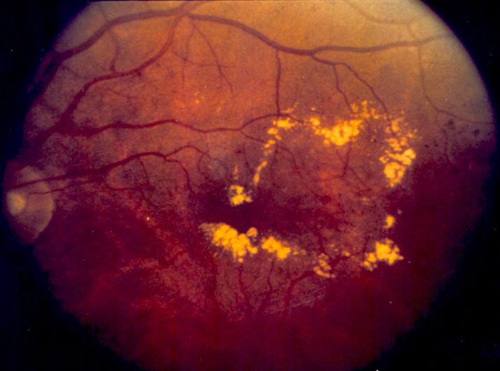
- Retinopathy affects 9-16% of people with type 2 diabetes and 24-27% of those with type 1, as reported in a German national guideline [S2].
- Risk factors include diabetes duration, the degree of hyperglycaemia, hypertension and diabetic nephropathy [S2].
- DMO results from leakage from diseased retinal capillaries, with thickening of the central retina and poor central vision [S6].
Confirm it
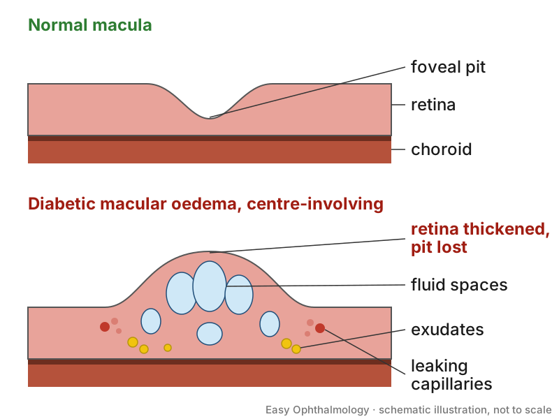
- The key treatment decision is whether the oedema involves the fovea (centre of the macula): intravitreal drugs are recommended for clinically significant oedema with foveal involvement [S2] [S3].
- Trials measured central subfield thickness on OCT; YOSEMITE and RHINE defined absence of DMO as central subfield thickness below 325 µm [S8].
- Baseline OCT also gives prognostic information (see Red flags) [S10]. No biomarker reached moderate certainty for predicting better vision [S10].
- Document findings in a standard way so the ophthalmologist and the diabetes or primary care team share them [S2].
Manage it
Before acting on this section, check the national and local guidelines, product information and regulatory approvals that apply where you practise. See the medical disclaimer.
Tier A (primary care / no specialist)
- Arrange risk-adapted eye examinations: every 2 years at low risk and every year for others, sooner as retinopathy becomes more severe [S2].
- Improve the general risk profile: glycaemia, blood pressure and kidney disease [S2].
- Explain the disease and the purpose of repeated injections; patient understanding was an enabler of adherence [S5].
- Trace patients who miss treatment visits [S5] [S4].
Tier B (general eye clinic)
- Macular laser. Compared with no treatment, laser reduced the chance of losing vision at 1 year (risk ratio 0.42) and increased resolution of clinically significant DMO at 3 years (risk ratio 1.55) [S3]. It remains an option for milder or non-central DMO [S3]. Subthreshold (especially micropulse) laser may be as effective as standard laser (low-certainty evidence) [S3].
- Bevacizumab first. In Protocol AC, 1.25 mg bevacizumab first, with a switch to 2.0 mg aflibercept if criteria were met (doses as reported in [S7]), gave a mean gain of 14.0 letters vs 15.0 letters with aflibercept alone over 2 years (difference 0.8 letters, 95% CI -0.9 to 2.5); 70% of bevacizumab-first eyes were switched [S7]. This trial enrolled eyes with vision 20/50 to 20/320 [S7].
- Steroids where anti-VEGF is unsuitable. The dexamethasone 0.7 mg implant improved vision slightly more than sham (mean difference -0.08 logMAR) but slightly less than anti-VEGF at 12 months (mean difference 0.07 logMAR), with about a fourfold increase in cataract progression and in pressure-lowering treatment [S6]. In pseudophakic eyes, cataract is not a concern [S6]. Weigh any benefit against pressure rise and the need for pressure-lowering treatment [S6]. Dosing beyond these reported values is not covered: follow local formularies.
Tier C (specialist centre)
- Choice of anti-VEGF. In Protocol T (2015), aflibercept 2.0 mg, bevacizumab 1.25 mg and ranibizumab 0.3 mg (as reported in [S1]) improved vision by 13.3, 9.7 and 11.2 letters at 1 year. With baseline vision 20/32 to 20/40 the drugs did not differ; at 20/50 or worse aflibercept gained more [S1]. A 2023 Cochrane network meta-analysis found small, possibly clinically insignificant 12-month advantages for aflibercept, brolucizumab and faricimab over ranibizumab, and no clinically important differences at 24 months [S9].
- Longer-acting regimens. Faricimab 6.0 mg treat-and-extend (up to every 16 weeks) matched aflibercept 2.0 mg every 8 weeks for vision at 2 years with fewer injections (median 10 and 11 vs 14); more than 60% of treat-and-extend patients were on 16-weekly dosing at week 96 [S8]. Aflibercept 8 mg every 12 or 16 weeks gave gains similar to aflibercept 2 mg every 8 weeks at 96 weeks (+8.8 and +7.5 vs +8.4 letters) with fewer injections (9.5 and 7.8 vs 13.8) [S11]. Both were industry-funded noninferiority trials [S8] [S11].
- Long-term follow-up. At 5 years in Protocol T, mean vision remained 7.4 letters above baseline, and 68% of eyes needed further injections between years 2 and 5 [S4].
- Steroid implants. The fluocinolone 0.19 mg implant improved vision slightly vs sham, but cataract progression occurred in about 8 in 10 participants [S6].
Refer when
| Situation | Urgency |
|---|---|
| Person with diabetes reports reduced or distorted central vision | Within a week to an eye clinic with OCT [EIC expert opinion] |
| Pain, redness or worsening vision after an intravitreal injection | Same-day, immediate review |
| DMO not involving the centre, vision unaffected | Routine, for assessment and laser; timing per your local service or specialist advice |
| Diabetes with no or mild retinopathy | Routine examination every 1 to 2 years by risk [S2] |
Pitfalls
- Assuming aflibercept is needed for every patient: with mild visual loss the three drugs did not differ [S1], and bevacizumab first showed no significant difference in 2-year vision [S7].
- Stopping follow-up after the first 2 years: vision drifted down between years 2 and 5 [S4].
- Using steroids in phakic eyes without planning for cataract and pressure monitoring [S6].
- Treating real-world results as trial results: under-treatment is common outside trials [S9] [S5].
Evidence notes
| Source | Date | Design |
|---|---|---|
| [S1] | 2015 | RCT (Protocol T), 660 adults, NIH-funded |
| [S2] | 2016 | National practice guideline (Germany) |
| [S3] | 2018 | Cochrane systematic review, 24 RCTs |
| [S4] | 2020 | Cohort follow-up of an RCT, 68% retained |
| [S5] | 2020 | Systematic review, 7 studies |
| [S6] | 2020 | Cochrane systematic review, 10 RCTs |
| [S7] | 2022 | RCT (Protocol AC), 312 eyes, NIH-funded |
| [S8] | 2024 | Two phase 3 noninferiority RCTs, industry-funded |
| [S9] | 2023 | Cochrane network meta-analysis, 23 RCTs |
| [S10] | 2026 | Systematic review and meta-analysis, 27 studies |
| [S11] | 2026 | Phase 2/3 noninferiority RCT |
- 13 of 23 trials in the Cochrane network meta-analysis had industry funding, and mortality estimates were very imprecise [S9].
- Most steroid trials had unclear or high risk of bias, and steroid vs anti-VEGF results were inconsistent [S6]. Laser trials were poorly reported; only one came from Africa [S3].
- In Protocol AC, serious adverse events were more frequent with aflibercept monotherapy (52% vs 36%) [S7]; the abstract does not attribute this to the drug.
- Not covered by these sources: drug cost and access, biosimilars, injection technique, vitrectomy for DMO, combination therapy, and DMO after cataract surgery.
Related articles
- Proliferative Diabetic Retinopathy: Laser, Anti-VEGF and Vitrectomy
- Anti-VEGF biosimilars and off-label bevacizumab
- OCT interpretation: macula and retinal nerve fibre layer
- Diabetic retinopathy screening programmes
References
See the numbered source list below. Each marker [S#] links to its PubMed and DOI record.
Sources
- [S1] Diabetic Retinopathy Clinical Research Network, Wells JA, Glassman AR, Ayala AR, Jampol LM, Aiello LP, et al. Aflibercept, bevacizumab, or ranibizumab for diabetic macular edema. The New England journal of medicine. (2015). PubMed 25692915 · doi:10.1056/nejmoa1414264
- [S2] Schorr SG, Hammes HP, Müller UA, Abholz HH, Landgraf R, Bertram B The Prevention and Treatment of Retinal Complications in Diabetes. Deutsches Arzteblatt international. (2016). PubMed 28073426 · doi:10.3238/arztebl.2016.0816
- [S3] Jorge EC, Jorge EN, Botelho M, Farat JG, Virgili G, El Dib R Monotherapy laser photocoagulation for diabetic macular oedema. The Cochrane database of systematic reviews. (2018). PubMed 30320466 · doi:10.1002/14651858.cd010859.pub2
- [S4] Glassman AR, Wells JA, Josic K, Maguire MG, Antoszyk AN, Baker C, et al. Five-Year Outcomes after Initial Aflibercept, Bevacizumab, or Ranibizumab Treatment for Diabetic Macular Edema (Protocol T Extension Study). Ophthalmology. (2020). PubMed 32402554 · doi:10.1016/j.ophtha.2020.03.021
- [S5] Rose MA, Vukicevic M, Koklanis K Adherence of patients with diabetic macular oedema to intravitreal injections: A systematic review. Clinical & experimental ophthalmology. (2020). PubMed 32829485 · doi:10.1111/ceo.13845
- [S6] Rittiphairoj T, Mir TA, Li T, Virgili G Intravitreal steroids for macular edema in diabetes. The Cochrane database of systematic reviews. (2020). PubMed 33206392 · doi:10.1002/14651858.cd005656.pub3
- [S7] Jhaveri CD, Glassman AR, Ferris FL, Liu D, Maguire MG, Allen JB, et al. Aflibercept Monotherapy or Bevacizumab First for Diabetic Macular Edema. The New England journal of medicine. (2022). PubMed 35833805 · doi:10.1056/nejmoa2204225
- [S8] Wong TY, Haskova Z, Asik K, Baumal CR, Csaky KG, Eter N, et al. Faricimab Treat-and-Extend for Diabetic Macular Edema: Two-Year Results from the Randomized Phase 3 YOSEMITE and RHINE Trials. Ophthalmology. (2024). PubMed 38158159 · doi:10.1016/j.ophtha.2023.12.026
- [S9] Virgili G, Curran K, Lucenteforte E, Peto T, Parravano M Anti-vascular endothelial growth factor for diabetic macular oedema: a network meta-analysis. The Cochrane database of systematic reviews. (2023). PubMed 38275741 · doi:10.1002/14651858.cd007419.pub7
- [S10] Nanji K, Grad J, Hatamnejad A, El-Sayes A, Mihalache A, Gemae M, et al. Baseline OCT Biomarkers Associated with Visual Acuity in Diabetic Macular Edema: A Systematic Review and Meta-analysis. Ophthalmology. (2026). PubMed 40803536 · doi:10.1016/j.ophtha.2025.07.038
- [S11] Do DV, Wykoff CC, Sivaprasad S, Brown DM, Boyer DS, Sakamoto T, et al. Intravitreal Aflibercept 8 mg for Diabetic Macular Edema: Ninety-Six-Week Results from the Randomized Phase 2/3 PHOTON Trial. Ophthalmology. (2026). PubMed 41223900 · doi:10.1016/j.ophtha.2025.10.028