Educational content only. Not personalised medical advice. Clinical decisions remain the responsibility of the treating clinician.
Proliferative Diabetic Retinopathy: Laser, Anti-VEGF and Vitrectomy
Proliferative diabetic retinopathy (PDR) is an advanced, sight-threatening stage of diabetic retinopathy in which abnormal new vessels grow on the retina, optic disc or anterior segment [S3], with a high risk of vitreous haemorrhage and tractional retinal detachment [S11]. Panretinal photocoagulation (PRP) remains the established treatment [S11] [S1]; anti-VEGF injections are an alternative or adjunct with similar long-term vision but a heavier visit burden and higher cost [S4] [S10].
Key points - PRP reduced the risk of severe visual loss by over 50% at 12 months compared with no or deferred treatment [S1]. - Ranibizumab was noninferior to PRP for vision at 2 years, and vision was similar (about 20/25) in both groups at 5 years [S2] [S4]. - Anti-VEGF caused less macular oedema and less visual field loss than PRP, but needed far more injections (mean 19.2 over 5 years) [S4]; its visual benefit over PRP is not clinically meaningful [S11] [S7]. - Most cost-effectiveness studies, largely from a US perspective, judged that anti-VEGF's extra cost outweighs its limited benefit over PRP [S10]. - Tractional detachment needs vitrectomy; earlier surgery with better preoperative vision gave better final vision [S6].
Evidence last checked: 2026-10-17 (PubMed abstracts).
Red flags
- Vitreous haemorrhage with vision loss in a person with diabetes: a complication of PDR [S7] [S5].
- Tractional retinal detachment, occurring in approximately 5% of people with PDR [S6]. Final vision after surgery is often poor (mean 0.94 logMAR, about 6/53), and worse when preoperative vision is worse [S6].
- Patient unlikely to attend frequent visits. Anti-VEGF alone needs repeated injections; anticipated visit compliance, cost and visit frequency should guide the choice of treatment [S4].
- High loss to follow-up. In Protocol S, only 66% of surviving participants completed the 5-year visit [S4].
Recognise it
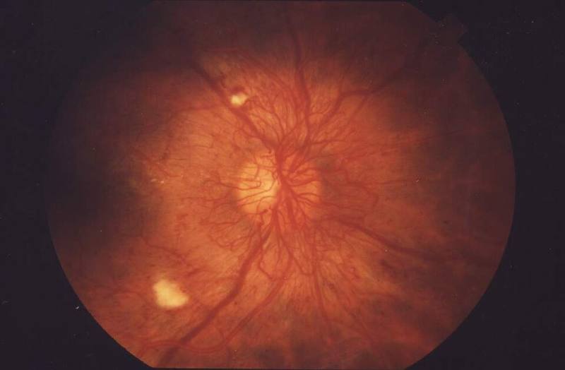
- PDR shows new vessels in the retina, optic nerve head or anterior segment [S3], and may present with vitreous haemorrhage [S7].
- Trial participants were mostly middle-aged (mean age 52 to 57 years) [S2] [S5] [S7]; mean HbA1c in the Cochrane review trials was 8.25% to 8.45% [S7].
- More intensive blood pressure control reduced the 5-year incidence of diabetic retinopathy (risk ratio 0.82, moderate certainty), particularly in hypertensive type 2 diabetes, but did not clearly slow progression or prevent PDR [S8].
Confirm it
- Trials judged activity as active or regressed neovascularisation [S2] and measured regression of new vessels [S7].
- When the view is blocked by vitreous haemorrhage, the abstracts do not cover the investigation of the retina behind it; follow local practice.
Manage it
Before acting on this section, check the national and local guidelines, product information and regulatory approvals that apply where you practise. See the medical disclaimer.
Tier A (primary care / no specialist)
- Recognise sudden vision loss or floaters in diabetes as possible vitreous haemorrhage and refer (see Refer when) [S5].
- Support blood pressure control, especially in hypertensive type 2 diabetes, for its modest effect on preventing retinopathy [S8]. More intensive control increased hypotension (risk ratio 2.04) [S8].
- Stress that PDR treatment needs follow-up: loss to follow-up was high even in trials [S4].
Tier B (general eye clinic)
- PRP is the mainstay. Laser reduced severe visual loss (risk ratio 0.46), progression of retinopathy (risk ratio 0.49) and vitreous haemorrhage (risk ratio 0.56) vs no or deferred treatment [S1]. In Protocol S, PRP was completed in 1 to 3 visits [S2].
- Laser type and pattern. Evidence comparing alternative lasers or patterns with standard ETDRS argon PRP is limited and mostly low or very low certainty [S3]. Diode laser was more painful than argon (risk ratio 3.12, moderate certainty) [S3].
- Pain relief. Analgesics or NSAIDs reduced pain during PRP compared with placebo in a meta-analysis of 13 RCTs; systemic analgesia was potentially better than eye drops [S9]. Specific doses are not covered; follow local formularies.
- Watch for macular oedema after PRP. Diabetic macular oedema developed in 28% of PRP eyes vs 9% of ranibizumab eyes at 2 years [S2].
Tier C (specialist centre)
- Anti-VEGF as alternative or adjunct. Ranibizumab 0.5 mg (as reported in [S2]) was noninferior to PRP at 2 years (+2.8 vs +0.2 letters), with less visual field loss, less vitrectomy (4% vs 15%) and less macular oedema [S2]. At 5 years, vision was similar (+3.1 vs +3.0 letters) [S4]. An individual participant data meta-analysis found a modest, not clinically meaningful, visual benefit at 1 year that declined over time, but fewer cases of macular oedema (relative risk 0.48) and retinal detachment (relative risk 0.41) [S11]. A Cochrane review found anti-VEGF with or without PRP probably reduced vitreous haemorrhage (risk ratio 0.72) and may reduce vitrectomy (risk ratio 0.67) [S7].
- Vitreous haemorrhage. In Protocol AB, initial aflibercept (4 monthly injections) vs vitrectomy with PRP gave no significant difference in vision over 24 weeks; vision recovered faster with vitrectomy at 4 weeks and was similar (about 20/40) at 2 years [S5]. About a third of each group later crossed over [S5]. The trial may have been underpowered to show a benefit of early vitrectomy [S5].
- Perioperative anti-VEGF. A network meta-analysis of 30 RCTs (2,646 eyes) found that anti-VEGF before or during vitrectomy improved postoperative vision and reduced intraoperative bleeding and recurrent haemorrhage, but evidence quality varied [S12].
- Tractional detachment. Vitrectomy failed to reattach the retina after one operation in 5.9% of eyes across 38 studies; preoperative vision was the only factor linked to final vision [S6].
Refer when
| Situation | Urgency |
|---|---|
| New vessels seen on the disc or retina in a person with diabetes | Urgent to an eye clinic able to perform PRP; timing per your local service or specialist advice |
| Sudden vision loss or new floaters in a person with diabetes (possible vitreous haemorrhage) | Urgent, within days [G2] |
| Suspected tractional detachment threatening the macula | Urgent to a vitreoretinal surgeon [S6]; timing per your local service or specialist advice |
| Pain, redness or worsening vision after an intravitreal injection | Same-day, immediate review [G1] |
Pitfalls
- Choosing anti-VEGF alone for a patient who cannot return regularly [S4].
- Expecting anti-VEGF to give clearly better vision than PRP: the difference is not clinically meaningful [S11] [S7].
- Forgetting macular oedema and field loss after PRP [S2] [S4].
- Delaying surgery for tractional detachment until vision has fallen [S6].
Evidence notes
| Source | Date | Design |
|---|---|---|
| [S1] | 2014 | Cochrane systematic review, 5 RCTs |
| [S2] | 2015 | RCT (Protocol S), 394 eyes, NIH-funded |
| [S3] | 2018 | Cochrane systematic review, 11 small RCTs |
| [S4] | 2018 | RCT (Protocol S), 5-year results |
| [S5] | 2020 | RCT (Protocol AB), 205 eyes |
| [S6] | 2023 | Systematic review and meta-analysis, 38 studies |
| [S7] | 2023 | Cochrane systematic review, 23 RCTs |
| [S8] | 2023 | Cochrane systematic review, 29 RCTs |
| [S9] | 2024 | Systematic review and meta-analysis, 13 RCTs |
| [S10] | 2025 | Systematic review of cost-effectiveness, NIHR-funded |
| [S11] | 2025 | Individual participant data meta-analysis, 12 RCTs, NIHR-funded |
| [S12] | 2026 | Network meta-analysis, 30 RCTs |
- The landmark laser trials are old, mostly from the USA, with performance and attrition bias [S1]. Most anti-VEGF RCTs had unclear or high risk of bias [S7].
- Protocol S was run at US sites and Protocol AB at US and Canadian sites [S2] [S5]; cost-effectiveness evidence was mostly from a US perspective and its transfer to other settings is unclear [S10].
- Long-term effectiveness and safety of anti-VEGF for PDR are unclear, as further PRP and injections are needed over time [S11].
- 8 of 29 blood pressure trials were sponsored entirely by pharmaceutical companies [S8].
- Not covered by these sources: PRP settings and number of burns, B-scan ultrasound, neovascular glaucoma, PDR in pregnancy, and screening programme design.
Related articles
- Diabetic Macular Oedema
- Diabetic retinopathy screening programmes
- Fluorescein and indocyanine green angiography
References
See the numbered source list below. Each marker [S#] links to its PubMed and DOI record.
Sources
- [S1] Evans JR, Michelessi M, Virgili G Laser photocoagulation for proliferative diabetic retinopathy. The Cochrane database of systematic reviews. (2014). PubMed 25420029 · doi:10.1002/14651858.cd011234.pub2
- [S2] Writing Committee for the Diabetic Retinopathy Clinical Research Network, Gross JG, Glassman AR, Jampol LM, Inusah S, Aiello LP, et al. Panretinal Photocoagulation vs Intravitreous Ranibizumab for Proliferative Diabetic Retinopathy: A Randomized Clinical Trial. JAMA. (2015). PubMed 26565927 · doi:10.1001/jama.2015.15217
- [S3] Moutray T, Evans JR, Lois N, Armstrong DJ, Peto T, Azuara-Blanco A Different lasers and techniques for proliferative diabetic retinopathy. The Cochrane database of systematic reviews. (2018). PubMed 29543992 · doi:10.1002/14651858.cd012314.pub2
- [S4] Gross JG, Glassman AR, Liu D, Sun JK, Antoszyk AN, Baker CW, et al. Five-Year Outcomes of Panretinal Photocoagulation vs Intravitreous Ranibizumab for Proliferative Diabetic Retinopathy: A Randomized Clinical Trial. JAMA ophthalmology. (2018). PubMed 30043039 · doi:10.1001/jamaophthalmol.2018.3255
- [S5] Antoszyk AN, Glassman AR, Beaulieu WT, Jampol LM, Jhaveri CD, Punjabi OS, et al. Effect of Intravitreous Aflibercept vs Vitrectomy With Panretinal Photocoagulation on Visual Acuity in Patients With Vitreous Hemorrhage From Proliferative Diabetic Retinopathy: A Randomized Clinical Trial. JAMA. (2020). PubMed 33320223 · doi:10.1001/jama.2020.23027
- [S6] McCullough P, Mohite A, Virgili G, Lois N Outcomes and Complications of Pars Plana Vitrectomy for Tractional Retinal Detachment in People With Diabetes: A Systematic Review and Meta-analysis. JAMA ophthalmology. (2023). PubMed 36633878 · doi:10.1001/jamaophthalmol.2022.5817
- [S7] Martinez-Zapata MJ, Salvador I, Martí-Carvajal AJ, Pijoan JI, Cordero JA, Ponomarev D, et al. Anti-vascular endothelial growth factor for proliferative diabetic retinopathy. The Cochrane database of systematic reviews. (2023). PubMed 36939655 · doi:10.1002/14651858.cd008721.pub3
- [S8] Do DV, Han G, Abariga SA, Sleilati G, Vedula SS, Hawkins BS Blood pressure control for diabetic retinopathy. The Cochrane database of systematic reviews. (2023). PubMed 36975019 · doi:10.1002/14651858.cd006127.pub3
- [S9] Arruda MP, Lima RV, Hira S, Moreira JLML, Barbosa LIT, Zinher MT, et al. A Comprehensive Meta-Analysis on the Role of Analgesics and Anti-Inflammatories in Pan-Retinal Photocoagulation. American journal of ophthalmology. (2024). PubMed 38942228 · doi:10.1016/j.ajo.2024.06.018
- [S10] Hodgson R, Walton M, Fulbright H, Bojke L, Walker R, Llewellyn A, et al. A systematic review of the cost-effectiveness of anti-VEGF drugs for the treatment of diabetic retinopathy. Health technology assessment (Winchester, England). (2025). PubMed 39881630 · doi:10.3310/nhyk3694
- [S11] Simmonds M, Llewellyn A, Walker R, Fulbright H, Walton M, Hodgson R, et al. Anti-VEGF drugs compared with laser photocoagulation for the treatment of proliferative diabetic retinopathy: a systematic review and individual participant data meta-analysis. Health technology assessment (Winchester, England). (2025). PubMed 40186529 · doi:10.3310/mjyp6578
- [S12] Zhao HQ, Zhou WD, Shao L, Zhang RH, Yang YH, Dong L, et al. Perioperative anti-VEGF therapy for proliferative diabetic retinopathy: a network meta-analysis of RCTs. Canadian journal of ophthalmology. Journal canadien d'ophtalmologie. (2026). PubMed 41620203 · doi:10.1016/j.jcjo.2026.01.006
Guidelines
- [G1] NHS Scotland (Community Eyecare Guidelines, eyes.nhs.scot). 7.4 Endophthalmitis, Post-Operative, (Exogenous Endophthalmitis) (2026). https://www.eyes.nhs.scot/eyecare-guidelines/section-7-uvea/74-endophthalmitis-post-operative-exogenous-endophthalmitis/
- [G2] NHS Scotland (Community Eyecare Guidelines, eyes.nhs.scot). 9.2 Vitreous Haemorrhage, No Previous Pan-Retinal Laser (n.d.). https://www.eyes.nhs.scot/eyecare-guidelines/section-9-vitreous/92-vitreous-haemorrhage-no-previous-pan-retinal-laser/