Educational content only. Not personalised medical advice. Clinical decisions remain the responsibility of the treating clinician.
Tonometry: Goldmann, rebound and home monitoring
Intraocular pressure (IOP) is the most important modifiable risk factor for glaucoma and fluctuates considerably within patients over short and long periods [S10]. Goldmann applanation tonometry (GAT) remains the reference standard but needs local anaesthetic, is difficult in children and poorly cooperative patients, and is influenced by corneal parameters [S9]. Rebound tonometers, including devices for self-measurement at home, widen access [S2] [S7] [S11].
Key points - No tonometer is perfect; a single IOP reading is an often error-prone snapshot and should be interpreted in the full clinical context [S2]. - Thicker corneas give artefactually high and thinner corneas artefactually low readings with GAT and many alternative tonometers [S6]; no adequately validated correction algorithm for GAT exists [S4]. - GAT underestimates IOP after laser refractive surgery [S1]. - Rebound tonometry needs no anaesthetic and is particularly useful in children [S2]; readings from different tonometers are often not interchangeable [S6]. - Tonometers can transmit infectious agents in vitro, and the best disinfection method is uncertain [S5].
Evidence last checked: 2026-10-17 (PubMed abstracts).
Red flags: findings that need urgent action
- Markedly raised IOP with a painful red eye, corneal oedema or reduced vision. Possible acute angle closure or other acute glaucoma: needs same-day eye assessment [G1].
- Corneal oedema in a child. Tono-Pen read 8.4 mm Hg higher on average than Icare PRO in oedematous eyes; confirm with a second method [S3].
- A "normal" reading after refractive surgery in a patient with a suspicious disc or field. GAT underestimates IOP after refractive surgery [S1].
- Low office IOP with progressing disease. Peak IOP often occurs outside office hours [S10].
Recognise it: what affects the reading
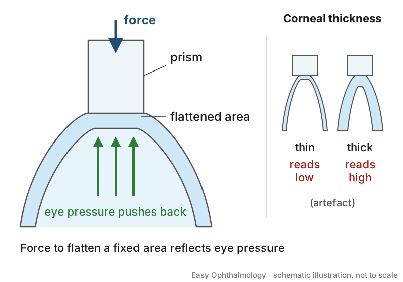
- Cornea. Variations in central corneal thickness (CCT) cause tonometry artefacts [S4]; demographics, environment, glaucoma treatment and the measuring device all influence CCT [S4]. Corneal biomechanical properties may be more strongly associated with glaucoma than CCT [S4].
- Children. In a cross-sectional study of 345 eyes of normal Egyptian children aged 2 months to 12 years (Perkins applanation), IOP was 5 to 20 mm Hg (mean 11.5 ± 2.34), rising with age and CCT (about 1 mm Hg per 100 µm) [S8]. This mean was lower than reported in other populations [S8].
- Anaesthesia. Induction and anaesthetic agents affect IOP; office IOPs in awake children are always the most accurate, according to one review [S6].
- Time of day. Glaucomatous eyes show greater IOP fluctuation than healthy eyes, and fluctuation is associated with progression independent of mean office IOP [S10].
Confirm it: choosing the tonometer
| Device | Strengths | Limits |
|---|---|---|
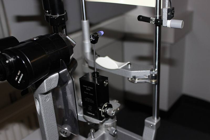
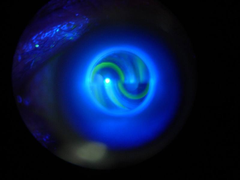
| Goldmann applanation | Reference standard [S9] | Needs anaesthetic, slit lamp, cooperation; corneal effects [S9] |
| Perkins (handheld applanation) | Used supine or under anaesthesia in children [S8] | Applanation corneal effects [S6] |
| Rebound (Icare) | No anaesthetic; useful in children [S2]; sitting or supine [S3] | Not interchangeable with other devices [S6] |
| Tono-Pen | Supine use under anaesthesia [S3] | About 2.2 mm Hg higher than Icare PRO in anaesthetised children [S3] |
| Dynamic contour | Fairly cornea-independent [S2] | Needs technical skill [S2] |
| Transpalpebral | Claims to measure through the closed lid [S2] | Too unreliable for routine use in comparative studies [S2] |
Manage it: measuring and acting on IOP at each tier
Before acting on this section, check the national and local guidelines, product information and regulatory approvals that apply where you practise. See the medical disclaimer.
Tier A (primary care / no specialist)
- Where a rebound tonometer is available, it needs no anaesthetic and suits children [S2]. Record the device used, because readings are often not interchangeable [S6].
- Interpret any single reading cautiously and alongside symptoms, the disc and acuity [S2].
- Disinfect the tonometer tip between patients following local infection-control policy; evidence on the most effective method is limited [S5].
Tier B (general eye clinic)
- Use GAT as the reference measurement [S9]. Measure CCT where possible and interpret IOP with it, without relying on a correction formula [S4].
- After refractive surgery, consider a less cornea-dependent device such as dynamic contour tonometry or Tono-Pen [S1].
- For children unable to cooperate at the slit lamp, use rebound or handheld applanation devices [S2] [S8], and compare with age-appropriate normal values [S8].
Tier C (specialist centre)
- For patients progressing despite apparently controlled office IOP, consider IOP measurement outside office hours [S10].
- Home rebound tonometers (iCare HOME, HOME2) allow anaesthesia-free self-measurement with good agreement with GAT and capture diurnal variation [S11]. Use them cautiously when CCT or IOP lie outside the validated range [S7].
- Contact lens sensors and implantable telemetric systems record continuous IOP; some have only animal data or pending long-term validation [S12].
- Barriers to home monitoring include cost, regulation, validation, compliance and workflow integration [S11] [S12]. RCTs are still needed to show that remote tonometry reduces vision loss [S10].
Refer when
| Situation | Urgency |
|---|---|
| Raised IOP with pain, red eye, corneal oedema or reduced vision | Same-day [G1] |
| Raised IOP in a child, or corneal enlargement or oedema in an infant | Urgent, within days [EIC expert opinion] |
| Raised IOP without symptoms, discs not yet assessed | Routine to an eye clinic able to assess discs and fields; timing per your local service or specialist advice |
| Glaucoma progressing at apparently normal office IOP [S10] | Routine specialist review; timing per your local service or specialist advice |
Pitfalls
- Treating one reading as the patient's true IOP [S2] [S10].
- Comparing readings from different devices as if equivalent [S6].
- Applying a CCT correction formula as if validated [S4].
- Missing raised IOP after refractive surgery [S1].
- Relying on transpalpebral readings [S2].
- Trusting Tono-Pen in corneal oedema [S3].
Evidence notes
| Source | Date | Design |
|---|---|---|
| [S1] | 2014 | Narrative review |
| [S2] | 2015 | Narrative review |
| [S3] | 2015 | Prospective comparative study, 100 eyes, randomised instrument order |
| [S4] | 2017 | Narrative review |
| [S5] | 2018 | Systematic review, 11 studies, mostly in vitro |
| [S6] | 2019 | Narrative review |
| [S7] | 2020 | Narrative review of a single device |
| [S8] | 2022 | Cross-sectional study with comparative meta-analysis |
| [S9] | 2021 | Narrative review |
| [S10] | 2024 | Narrative review |
| [S11] | 2026 | Narrative review |
| [S12] | 2026 | Narrative review |
- The evidence base on this sheet is thin: mostly narrative reviews, with one small comparative study [S3] and one cross-sectional normative study [S8]. No systematic review of tonometer agreement was available.
- The paediatric normal range comes from one Egyptian population and was lower than elsewhere [S8].
- Infection transmission evidence is largely in vitro [S5].
- The search returned many off-topic records (sleep apnoea and arterial tonometry, blood pressure monitors).
- Not covered by these sources: step-by-step GAT technique and calibration checks, numeric agreement limits between devices in adults, IOP thresholds for treatment, and specific disinfection protocols.
Related articles
References
See the numbered source list below. Each marker [S#] links to its PubMed and DOI record.
Sources
- [S1] Yao WJ, Crossan AS An update on postrefractive surgery intraocular pressure determination. Current opinion in ophthalmology. (2014). PubMed 24807062 · doi:10.1097/icu.0000000000000065
- [S2] Okafor KC, Brandt JD Measuring intraocular pressure. Current opinion in ophthalmology. (2015). PubMed 25594767 · doi:10.1097/icu.0000000000000129
- [S3] McKee EC, Ely AL, Duncan JE, Dosunmu EO, Freedman SF A comparison of Icare PRO and Tono-Pen XL tonometers in anesthetized children. Journal of AAPOS : the official publication of the American Association for Pediatric Ophthalmology and Strabismus. (2015). PubMed 26235790 · doi:10.1016/j.jaapos.2015.04.004
- [S4] Sng CC, Ang M, Barton K Central corneal thickness in glaucoma. Current opinion in ophthalmology. (2017). PubMed 27764022 · doi:10.1097/icu.0000000000000335
- [S5] Atkins N, Hodge W, Li B A Systematic Review Regarding Tonometry and the Transmission of Infectious Diseases. Journal of clinical medicine research. (2018). PubMed 29416571 · doi:10.14740/jocmr3294w
- [S6] Fayed MA, Chen TC Pediatric intraocular pressure measurements: Tonometers, central corneal thickness, and anesthesia. Survey of ophthalmology. (2019). PubMed 31132392 · doi:10.1016/j.survophthal.2019.05.003
- [S7] Liu J, De Francesco T, Schlenker M, Ahmed II Icare Home Tonometer: A Review of Characteristics and Clinical Utility. Clinical ophthalmology (Auckland, N.Z.). (2020). PubMed 33262568 · doi:10.2147/opth.s284844
- [S8] Moussa IR, Kassem RR, Edris NA, Khalil DH Normal intraocular pressure in Egyptian children and meta-analysis. Eye (London, England). (2022). PubMed 34145417 · doi:10.1038/s41433-021-01633-8
- [S9] Brusini P, Salvetat ML, Zeppieri M How to Measure Intraocular Pressure: An Updated Review of Various Tonometers. Journal of clinical medicine. (2021). PubMed 34501306 · doi:10.3390/jcm10173860
- [S10] Asrani SG, McGlumphy EJ, Al-Aswad LA, Chaya CJ, Lin S, Musch DC, et al. The relationship between intraocular pressure and glaucoma: An evolving concept. Progress in retinal and eye research. (2024). PubMed 39303763 · doi:10.1016/j.preteyeres.2024.101303
- [S11] Kamdar A, Swaminathan SS Beyond the Clinic: Home-Based Monitoring Strategies in Glaucoma Care. International ophthalmology clinics. (2026). PubMed 42312610 · doi:10.1097/iio.0000000000000628
- [S12] Mansoori T, Chaturvedi AK, Kong GY, Panigrahi A, Malik MA, Mansouri K, et al. Home monitoring devices for glaucoma. Indian journal of ophthalmology. (2026). PubMed 42809249 · doi:10.4103/ijo.ijo_84_26
Guidelines
- [G1] NHS Scotland Community Eyecare Guidelines. 6.4 Glaucoma, Acute Angle Closure (n.d.). https://www.eyes.nhs.scot/eyecare-guidelines/section-6-glaucoma/64-glaucoma-acute-angle-closure/