Educational content only. Not personalised medical advice. Clinical decisions remain the responsibility of the treating clinician.
Orbital fractures
Orbital fractures commonly result from blunt trauma such as accidents or assaults and make up a significant proportion of facial fractures [S11]. Orbital trauma can cause permanent vision loss, and the main early dangers are an associated globe injury, retrobulbar haematoma, muscle entrapment with ischaemia and orbital apex involvement [S12]. Many floor fractures heal without intervention [S8].
Key points - Always look for a concomitant eye injury; missing one is a feared consequence of acute fracture assessment [S10]. - CT is the primary imaging modality for fracture pattern, globe injury, muscle entrapment and vascular complications [S12]. - A paediatric trapdoor fracture with entrapment, marked loss of motility or an oculocardiac reflex should be repaired within 24 hours of diagnosis (expert recommendation) [S1]. - Fractures without entrapment, especially in children, can often be managed conservatively with close follow-up [S1] [S9]. - Delayed enophthalmos develops in 7 to 10% of conservatively managed fractures; predictors exist but the evidence is low level [S7].
Evidence last checked: 2026-10-17 (PubMed abstracts).
Red flags
- Globe rupture or retrobulbar haematoma found with the fracture: both require urgent intervention [S12].
- Muscle entrapment with ischaemia on CT [S12].
- "White-eyed" blowout fracture: often overlooked because it has few overt clinical and radiographic features, but it can cause significant morbidity [S3].
- Child with marked or complete loss of eye movement, or an oculocardiac reflex (trapdoor fracture) [S1].
- Orbital apex involvement [S12].
Recognise it
- Floor blowout fractures may cause disabling double vision and globe malposition [S1] [S11].
- Patients are often in pain and may have bleeding around the eye, which makes examination difficult [S5].
- Blowout fractures usually lie in the orbital floor medial to the infraorbital nerve and in the medial wall [S4].
- In children, a trapdoor fracture occurs when herniated tissue is trapped by a recoiled bone fragment [S1].
Confirm it
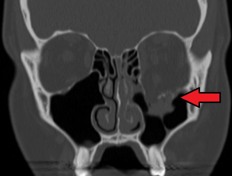
- Examine the eye itself: concomitant ocular injuries range in severity, and their sequelae may be serious [S10].
- CT: multidetector CT is the primary modality [S12]; CT with 3D reconstruction supports management [S11].
- CT findings: comminuted, hinged or linear fractures; herniation or incarceration of orbital fat at the fracture site; and sometimes orbital emphysema and haematoma [S4].
- Predictors of delayed enophthalmos on imaging include linear measurements, involvement of specific intraorbital structures, rounding of the inferior rectus, fracture area and volumetric analysis by fracture site [S7].
Manage it
Before acting on this section, check the national and local guidelines, product information and regulatory approvals that apply where you practise. See the medical disclaimer.
Tier A (primary care / no specialist)
- Many eyelid and orbital injuries require quick and immediate intervention; examine the eye and refer [S5].
- Refer every suspected orbital fracture for eye assessment and imaging (see table).
- An immediate ophthalmology review for every orbital fracture is debated because many examinations are normal and the burden on services is high [S10]. Patients with any red flag above need urgent specialist review [S12].
Tier B (general eye clinic)
- Examine for a concomitant ocular injury, restricted motility, diplopia and globe malposition [S1] [S10] [S12].
- Conservative care: many floor fractures heal without intervention or adverse sequelae [S8]. In children, most fractures can initially be followed to see whether disabling diplopia resolves [S1].
- Enophthalmos: in children, enophthalmos does not need to be prevented by early surgery; it can be allowed to develop and repaired later if noticeable [S1].
- Management is multidisciplinary [S11].
Tier C (specialist centre)
- Timing: data on the optimal timing of repair conflict, and indications remain controversial [S8] [S11]. The exception is the paediatric trapdoor fracture, for which repair within 24 hours of diagnosis is recommended [S1].
- Approach: in children, a simple transconjunctival incision is preferred to a cutaneous one, and the implant must sit on the posterior bony ledge; misplacement under the ledge is the most common reason for revision [S1].
- Outcomes in children: approximately 85% of children with preoperative diplopia recover completely after repair [S1].
- Implants: options range from autologous grafts to titanium mesh [S11]. Resorbable implants suit isolated floor or medial wall fractures with intact buttresses, not load-bearing areas; reported complications include enophthalmos (5%-16%), diplopia (0%-16%) and infraorbital nerve hypoaesthesia (2%-18%) [S2]. In children, resorbable implants are increasingly preferred [S9].
- Patient-specific implants and navigation can improve precision [S8] [S11], but outcomes with patient-specific implants are largely comparable to conventional implants [S6].
Refer when
| Situation | Urgency |
|---|---|
| Fracture with suspected globe rupture or retrobulbar haematoma | Immediate [S12] |
| Child with entrapment, marked motility loss or oculocardiac reflex | Immediate; repair within 24 hours of diagnosis [S1] |
| Fracture with reduced vision or an abnormal eye examination | Same-day ophthalmology review |
| Suspected fracture without red flags | Emergency referral for eye examination and CT [EIC expert opinion] |
| Persistent diplopia or noticeable enophthalmos after the swelling settles | Routine, within weeks, to an orbital or maxillofacial surgeon [S1] [S7] |
Pitfalls
- Missing a "white-eyed" blowout fracture, which has few overt clinical and radiographic signs [S3].
- Focusing on the bone and missing a globe injury [S10].
- Operating early to prevent enophthalmos in children when it can be watched [S1].
- Assuming custom implants guarantee better outcomes [S6].
Evidence notes
| Source | Date | Design |
|---|---|---|
| [S1] | 2017 | Narrative review with guidance (paediatric) |
| [S2] | 2018 | Systematic review, 27 studies (resorbable implants) |
| [S3] | 2019 | Narrative review |
| [S4] | 2021 | Narrative review with institutional imaging |
| [S5] | 2020 | Narrative review (primary care) |
| [S6] | 2022 | Systematic review, 15 studies (patient-specific implants) |
| [S7] | 2022 | Systematic review, 16 studies (delayed enophthalmos) |
| [S8] | 2022 | Narrative review |
| [S9] | 2023 | Narrative review (paediatric) |
| [S10] | 2025 | Narrative review |
| [S11] | 2025 | Narrative review |
| [S12] | 2026 | Narrative review (imaging) |
- Low-level evidence throughout. There is a paucity of level I and II evidence [S7] and of prospective studies [S11]; there are no RCTs in this source set. The 24-hour trapdoor recommendation is expert opinion [S1].
- Paediatric orbital fractures are rare, and studies have small cohorts and short follow-up [S9].
- Not covered by these sources: nose-blowing advice, prophylactic antibiotics, systemic steroids, specific CT protocols and repair timing in adults.
Related articles
- Blunt eye injury and traumatic hyphaema
- Orbital compartment syndrome and retrobulbar haemorrhage
- Traumatic optic neuropathy
References
See the numbered source list below. Each marker [S#] links to its PubMed and DOI record.
Sources
- [S1] Chung SY, Langer PD Pediatric orbital blowout fractures. Current opinion in ophthalmology. (2017). PubMed 28797015 · doi:10.1097/icu.0000000000000407
- [S2] Ramesh S, Hubschman S, Goldberg R Resorbable Implants for Orbital Fractures: A Systematic Review. Annals of plastic surgery. (2018). PubMed 29851726 · doi:10.1097/sap.0000000000001504
- [S3] Saggese NP, Mohammadi E, Cardo VA The 'White-eyed' Orbital Blowout Fracture: An Easily Overlooked Injury in Maxillofacial Trauma. Cureus. (2019). PubMed 31245200 · doi:10.7759/cureus.4412
- [S4] Valencia MR, Miyazaki H, Ito M, Nishimura K, Kakizaki H, Takahashi Y Radiological findings of orbital blowout fractures: a review. Orbit (Amsterdam, Netherlands). (2021). PubMed 32212885 · doi:10.1080/01676830.2020.1744670
- [S5] Gordon AA, Tran LT, Phelps PO Eyelid and orbital trauma for the primary care physician. Disease-a-month : DM. (2020). PubMed 32622682 · doi:10.1016/j.disamonth.2020.101045
- [S6] Maher DI, Hall AJ, Gwini S, Ben Artsi E Patient-specific Implants for Orbital Fractures: A Systematic Review. Ophthalmic plastic and reconstructive surgery. (2022). PubMed 34750315 · doi:10.1097/iop.0000000000002089
- [S7] Choi A, Sisson A, Olson K, Sivam S Predictors of Delayed Enophthalmos After Orbital Fractures: A Systematic Review. Facial plastic surgery & aesthetic medicine. (2022). PubMed 35179991 · doi:10.1089/fpsam.2021.0177
- [S8] Patel S, Shokri T, Ziai K, Lighthall JG Controversies and Contemporary Management of Orbital Floor Fractures. Craniomaxillofacial trauma & reconstruction. (2022). PubMed 36081678 · doi:10.1177/19433875211026430
- [S9] Coviello C, Williams KJ, Sivam SK Pediatric orbital fractures. Current opinion in otolaryngology & head and neck surgery. (2023). PubMed 36976962 · doi:10.1097/moo.0000000000000887
- [S10] Webb KL, Oyer S, Park S Is Ophthalmology Consult Needed for Acute Orbital Fractures? The Laryngoscope. (2025). PubMed 40391877 · doi:10.1002/lary.32272
- [S11] Sawaf T, Justicz N, Vakharia KT Updates on the Evaluation and Treatment of Orbital Fractures. Facial plastic surgery clinics of North America. (2025). PubMed 40581448 · doi:10.1016/j.fsc.2025.03.002
- [S12] Hood CM, Gardiner MF, Bleicher ID, Lev MH, Jutras M Imaging, Management, and Treatment of Orbital Trauma. Radiologic clinics of North America. (2026). PubMed 42230064 · doi:10.1016/j.rcl.2026.03.001