Educational content only. Not personalised medical advice. Clinical decisions remain the responsibility of the treating clinician.
Giant Cell Arteritis
Giant cell arteritis (GCA) is a systemic inflammatory vasculitis mainly affecting people over 50 years [S9]. It is a medical emergency because of the risk of sudden, irreversible loss of sight and, more rarely, stroke [S4] [S9]. Immediate high-dose glucocorticoids are the first-line treatment, and glucocorticoid-sparing agents reduce relapse and steroid exposure [S1] [S2].
Key points - Start high-dose glucocorticoids immediately in active GCA; EULAR reports 40-60 mg/day prednisone-equivalent for induction [S2]. - Confirm the diagnosis by imaging or histology [S2]; EULAR now recommends ultrasound as the first-line imaging test, including the axillary arteries [S8]. - No single symptom or test confirms or excludes GCA; jaw claudication, temporal artery abnormalities, platelets above 400 × 10³/µL and very high ESR raise the likelihood [S5]. - Relapse occurs in almost half of patients, mainly in the first two years [S7]. - Tocilizumab (RCT) and upadacitinib 15 mg (RCT) improve sustained remission with a shorter steroid taper [S1] [S11].
Evidence last checked: 2026-10-17 (PubMed abstracts).
Red flags
- Any visual symptom in a patient over 50 with headache or jaw claudication: GCA threatens sudden irreversible sight loss [S4] [S9].
- Jaw claudication (positive likelihood ratio 4.90) and limb claudication (6.01) [S5].
- Temporal artery thickening, loss of pulse or tenderness (positive likelihood ratios 4.70, 3.25 and 3.14) [S5].
- Large vessel involvement, which predicts relapse [S7].
Recognise it
- In a meta-analysis of 68 studies (14,037 patients with suspected GCA), features that increased the probability of GCA included ESR above 100 mm/h (positive LR 3.11) and platelet count above 400 × 10³/µL (3.75) [S5].
- Features whose absence lowered the probability: ESR above 40 mm/h (negative LR 0.18), CRP of 2.5 mg/dL or more (0.38) and age over 70 years (0.48) [S5].
- Combinations of features help direct further tests such as vascular imaging or biopsy [S5].
- Headache is a cardinal symptom of cranial GCA [S4].
Confirm it
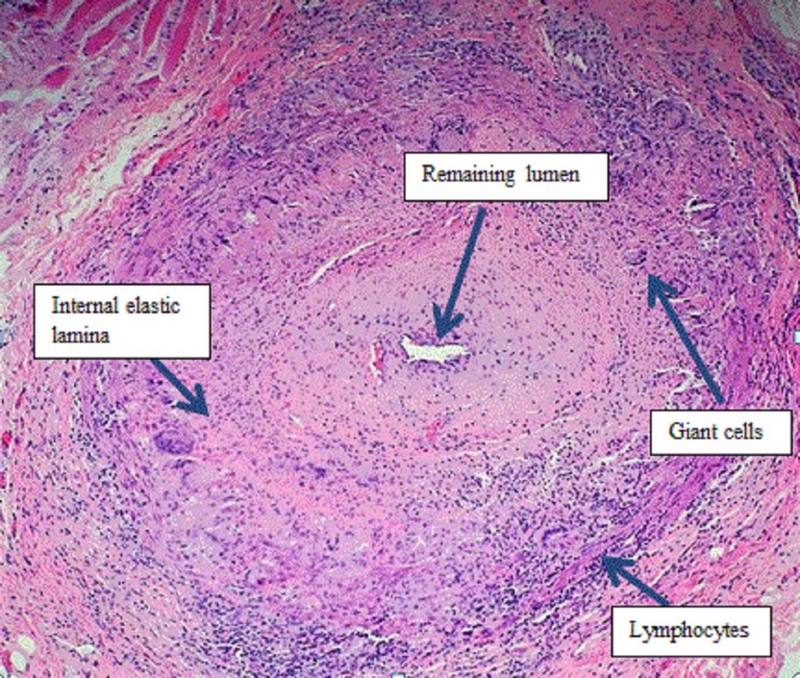
- Ultrasound is the first-line imaging test in suspected GCA; FDG-PET or MRI of cranial and extracranial arteries are alternatives [S8]. This is guideline advice based on evidence and expert opinion [S8].
- Temporal artery biopsy: pooled sensitivity 77.3% in 32 studies (3092 patients), with high heterogeneity, so a negative biopsy does not exclude GCA [S3].
- Ultrasound vs biopsy: a Cochrane review (2024) found very low-certainty evidence; ultrasound appears moderately sensitive when specificity is good, and no conclusion could be drawn on whether it can replace biopsy [S9].
- Fast-track pathways and advanced imaging help reduce diagnostic delay in Europe [S4].
Manage it
Before acting on this section, check the national and local guidelines, product information and regulatory approvals that apply where you practise. See the medical disclaimer.
Tier A (primary care / no specialist)
- Suspect GCA in anyone over 50 with headache [S4], jaw claudication, scalp or temporal tenderness, or visual symptoms [S5] [S9].
- Send ESR, CRP and platelet count if available [S5], but do not wait for results before arranging urgent treatment and referral [S2].
- Glucocorticoids should be started immediately in active GCA [S2]. If a clinician can prescribe, start as per local protocol; EULAR reports 40-60 mg/day prednisone-equivalent (as reported in [S2]).
Tier B (general eye clinic)
- Document visual function and start glucocorticoids immediately in active GCA [S2]; sight loss can be sudden and irreversible [S4].
- Arrange ultrasound or temporal artery biopsy without delaying treatment [S2] [S8]. Biopsy was positive in only 77% of confirmed cases [S3].
- Co-manage with rheumatology or internal medicine for long-term steroid tapering [S6] [S10].
Tier C (specialist centre)
- Glucocorticoid-sparing therapy: - Tocilizumab (GiACTA, 251 patients): sustained remission at 52 weeks in 56% (weekly) and 53% (every other week) vs 14% and 18% with placebo plus 26- or 52-week prednisone tapers; cumulative prednisone 1862 mg vs 3296 to 3818 mg [S1]. Dose as reported in [S1]: 162 mg subcutaneously. Industry-funded. - Upadacitinib (SELECT-GCA, 428 patients): sustained remission 46.4% with 15 mg daily vs 29.0% with placebo; 7.5 mg was not superior; no major adverse cardiovascular events [S11]. Industry-funded. - Methotrexate may be used as an alternative to tocilizumab [S2] [S10].
- Who should get tocilizumab: EULAR advises it in refractory or relapsing disease or high risk of glucocorticoid adverse effects [S2]. The French group (2025) recommends it at diagnosis after a major cardiovascular event, fracture with osteoporosis, psychiatric event with steroids, complicated diabetes, or previous steroid use over 6 months, for at least 12 months [S10]. The ACR guideline supports glucocorticoid-sparing agents [S6].
- Antiplatelet or anticoagulant therapy is no longer routinely recommended unless indicated for other reasons [S2].
- Relapse monitoring: cumulative relapse rates were 32% at 1 year, 44% at 2 years and 47% at 5 years; female sex and large vessel involvement predicted relapse [S7]. Imaging can help assess suspected relapse when inflammatory markers are unreliable [S8].
Low-resource note: where ultrasound, biopsy or biologics are unavailable, immediate glucocorticoids remain the first-line treatment [S2] [S4], and methotrexate is an alternative glucocorticoid-sparing agent [S2].
Refer when
| Situation | Urgency |
|---|---|
| Suspected GCA with visual symptoms or signs | Immediate: medical emergency; start glucocorticoids [S2] [S4] |
| Suspected GCA without visual symptoms | Same-day assessment and treatment [S2] [S4] |
| Relapse of symptoms or rising markers during taper | Urgent; timing per your local service or specialist advice [S7] |
| Long-term steroid use or steroid complications | Routine specialist review for steroid-sparing therapy; timing per your local service or specialist advice [S10] |
Pitfalls
- Delaying glucocorticoids while waiting for biopsy or imaging [S2].
- Excluding GCA on a negative biopsy [S3].
- Relying on one symptom or test [S5].
- Tapering too fast without a plan for relapse, which affects almost half of patients [S7].
Evidence notes
| Source | Date | Design |
|---|---|---|
| [S1] | 2017 | Phase 3 RCT (GiACTA), 251 patients; industry-funded |
| [S2] | 2020 | EULAR recommendations, systematic review and expert opinion |
| [S3] | 2020 | Meta-analysis, 32 studies |
| [S4] | 2020 | European Headache Federation recommendations |
| [S5] | 2020 | Diagnostic accuracy meta-analysis, 68 studies |
| [S6] | 2021 | ACR/VF guideline, GRADE; mostly conditional recommendations |
| [S7] | 2023 | Meta-analysis of relapse, observational |
| [S8] | 2024 | EULAR imaging recommendations, 2023 update |
| [S9] | 2024 | Cochrane diagnostic accuracy review, very low certainty |
| [S10] | 2025 | French national recommendations, expert consensus |
| [S11] | 2025 | Phase 3 RCT (SELECT-GCA); industry-funded |
- Conflict: EULAR recommends ultrasound first-line [S8], while the Cochrane review found very low-certainty evidence and could not conclude whether ultrasound can replace biopsy [S9]. Biopsy sensitivity estimates also vary widely [S3].
- The ACR guideline has only 2 strong recommendations; the rest are conditional because of low-quality evidence [S6].
- The tocilizumab and upadacitinib trials were industry-funded and compared against placebo with steroid tapers [S1] [S11].
- Not covered by these sources: intravenous methylprednisolone for visual loss, bone and gastric protection, the size of the visual-loss risk, and data from low- and middle-income countries.
Related articles
- Non-Arteritic Anterior Ischaemic Optic Neuropathy
- Central Retinal Artery Occlusion: An Ocular Stroke
- Sudden painless vision loss: triage
References
See the numbered source list below. Each marker [S#] links to its PubMed and DOI record.
Sources
- [S1] Stone JH, Tuckwell K, Dimonaco S, Klearman M, Aringer M, Blockmans D, et al. Trial of Tocilizumab in Giant-Cell Arteritis. The New England journal of medicine. (2017). PubMed 28745999 · doi:10.1056/nejmoa1613849
- [S2] Hellmich B, Agueda A, Monti S, Buttgereit F, de Boysson H, Brouwer E, et al. 2018 Update of the EULAR recommendations for the management of large vessel vasculitis. Annals of the rheumatic diseases. (2020). PubMed 31270110 · doi:10.1136/annrheumdis-2019-215672
- [S3] Rubenstein E, Maldini C, Gonzalez-Chiappe S, Chevret S, Mahr A Sensitivity of temporal artery biopsy in the diagnosis of giant cell arteritis: a systematic literature review and meta-analysis. Rheumatology (Oxford, England). (2020). PubMed 31529073 · doi:10.1093/rheumatology/kez385
- [S4] Mollan SP, Paemeleire K, Versijpt J, Luqmani R, Sinclair AJ European Headache Federation recommendations for neurologists managing giant cell arteritis. The journal of headache and pain. (2020). PubMed 32183689 · doi:10.1186/s10194-020-01093-7
- [S5] van der Geest KSM, Sandovici M, Brouwer E, Mackie SL Diagnostic Accuracy of Symptoms, Physical Signs, and Laboratory Tests for Giant Cell Arteritis: A Systematic Review and Meta-analysis. JAMA internal medicine. (2020). PubMed 32804186 · doi:10.1001/jamainternmed.2020.3050
- [S6] Maz M, Chung SA, Abril A, Langford CA, Gorelik M, Guyatt G, et al. 2021 American College of Rheumatology/Vasculitis Foundation Guideline for the Management of Giant Cell Arteritis and Takayasu Arteritis. Arthritis care & research. (2021). PubMed 34235871 · doi:10.1002/acr.24632
- [S7] Moreel L, Betrains A, Molenberghs G, Vanderschueren S, Blockmans D Epidemiology and predictors of relapse in giant cell arteritis: A systematic review and meta-analysis. Joint bone spine. (2023). PubMed 36410684 · doi:10.1016/j.jbspin.2022.105494
- [S8] Dejaco C, Ramiro S, Bond M, Bosch P, Ponte C, Mackie SL, et al. EULAR recommendations for the use of imaging in large vessel vasculitis in clinical practice: 2023 update. Annals of the rheumatic diseases. (2024). PubMed 37550004 · doi:10.1136/ard-2023-224543
- [S9] Pouncey AL, Yeldham G, Magan T, Lucenteforte E, Jaffer U, Virgili G Halo sign on temporal artery ultrasound versus temporal artery biopsy for giant cell arteritis. The Cochrane database of systematic reviews. (2024). PubMed 38323659 · doi:10.1002/14651858.cd013199.pub2
- [S10] de Boysson H, Devauchelle-Pensec V, Agard C, André M, Bienvenu B, Bonnotte B, et al. Use of immunosuppressants and biologics in giant cell arteritis: Recommendations of the French Study Group for Large Vessel Vasculitis (GEFA). La Revue de medecine interne. (2025). PubMed 39500637 · doi:10.1016/j.revmed.2024.10.006
- [S11] Blockmans D, Penn SK, Setty AR, Schmidt WA, Rubbert-Roth A, Hauge EM, et al. A Phase 3 Trial of Upadacitinib for Giant-Cell Arteritis. The New England journal of medicine. (2025). PubMed 40174237 · doi:10.1056/nejmoa2413449