Educational content only. Not personalised medical advice. Clinical decisions remain the responsibility of the treating clinician.
Ocular Motor Cranial Nerve Palsies
Palsies of the third (oculomotor), fourth (trochlear) and sixth (abducens) cranial nerves cause binocular diplopia and can result from ischaemia, inflammation, infection, compression or trauma [S2]. Most isolated palsies in people over 50 are microvascular, but aneurysm, tumour and giant cell arteritis must be considered [S3]. A third nerve palsy can signal a compressive aneurysm, which needs urgent vascular imaging [S5].
Key points - All patients with a third nerve palsy need urgent brain CT angiography to exclude a compressive aneurysm [S5]. - Microvascular ischaemia was the commonest cause in large series (26.5% of third and 36.7% of sixth nerve palsies), but neoplastic and vascular causes were frequent [S9] [S7]. - Imaging of isolated palsies in older adults is debated; one review recommends contrast MRI for all, while another notes that some clinicians observe sixth nerve palsy with vascular risk factors for 2 to 3 months [S2] [S5]. - More than one cranial nerve palsy, or brainstem symptoms, needs urgent neuroimaging [S5]. - Botulinum toxin helps acute palsies (success 79%) more than chronic ones (33%), in mostly observational data [S8].
Evidence last checked: 2026-10-17 (PubMed abstracts).
Red flags
- Any third nerve palsy: urgent CT angiography for aneurysm [S5]. In adults, a dilated pupil in third nerve palsy suggests compression; this does not apply in children [S1].
- Recent binocular diplopia with brainstem symptoms: urgent brain MRI [S5].
- Multiple cranial nerve palsies: urgent imaging, with attention to the cavernous sinus and superior orbital fissure [S5].
- Age 50 or older: consider aneurysm, malignancy and giant cell arteritis as well as microvascular ischaemia [S3].
- Sixth nerve palsy in a child: tumour risk may justify early neuroimaging [S1].
Recognise it
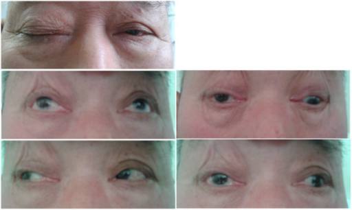
- Third nerve palsy: ptosis, diplopia and periorbital pain are common in diabetic palsies; pupil sparing is characteristic of the diabetic form [S11]. In 633 patients with isolated third nerve palsy (Korea), causes were microvascular 26.5%, vascular anomalies 17.4%, neoplastic 13.6%, inflammatory 12.5%, idiopathic 9.5% and traumatic 8.4% [S9].
- Sixth nerve palsy: in 807 patients, causes were microvascular 36.7%, idiopathic 17.7%, neoplastic 14.3%, vascular anomalies 10.2%, inflammatory 9.4% and traumatic 4.3% [S7]. The distribution varied with age, sex and the specialty managing the patient [S7] [S9].
- Fourth nerve palsy: hyperdeviation that worsens in downgaze should prompt contrast MRI [S5].
- Aneurysms can also cause non-oculomotor palsies; the sixth nerve was most often affected in a systematic review of 47 patients [S10].
- Children: aberrant regeneration is very common after congenital third nerve palsy, and Duane syndrome results from early repair after congenital sixth nerve palsy [S1].
- Mimics: suspect ocular myasthenia when misalignment fits no single nerve pattern; orbital disease, most often thyroid eye disease, restricts motility [S5].
Confirm it
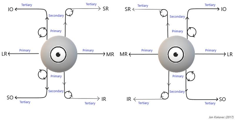
- Separate monocular from binocular diplopia: monocular diplopia, and comitant misalignment with full motility, need ophthalmology referral rather than further testing [S5].
- Examine neighbouring cranial nerves, including in children [S1].
- Imaging:
- Third nerve palsy: urgent CT angiography [S5].
- Sixth nerve palsy: contrast MRI, except that some clinicians observe patients over 50 with microvascular risk factors, most of whom improve in 2 to 3 months [S5].
- A prospective study cited in a review found structural lesions on MRI in 16.5% of adults with acute ocular motor mononeuropathy, and in 4.6% of those with fourth or sixth nerve palsies and no risk factors; the reviewers recommend contrast MRI for all isolated acute palsies regardless of age [S2].
Manage it
Before acting on this section, check the national and local guidelines, product information and regulatory approvals that apply where you practise. See the medical disclaimer.
Tier A (primary care / no specialist)
- A new third nerve palsy, or diplopia with other neurological signs, needs urgent transfer for vascular or brain imaging [S5].
- Check for diabetes and vascular risk factors: optimised glycaemic and vascular risk-factor control is part of treatment for diabetic palsies [S11].
Tier B (general eye clinic)
- Document the pattern of deviation, ptosis and pupils [S5] [S11].
- Arrange imaging according to the nerve involved [S2] [S5]; consider giant cell arteritis in patients over 50 [S3].
- For presumed microvascular sixth nerve palsy managed by observation, expect improvement within 2 to 3 months; image if it does not improve [S5].
- Spontaneous recovery is seen in many diabetic palsies [S11].
Tier C (specialist centre)
- Botulinum toxin: a meta-analysis of 38 mainly observational studies (643 patients) found success in 79% of acute and 33% of chronic palsies; odds ratio vs expectant management 2.67 for acute palsy and 0.87 for chronic palsy [S8]. A Cochrane review found one small RCT (47 people, acute sixth nerve palsy) with low-certainty evidence of a higher full recovery rate (risk ratio 1.19, 95% CI 0.96 to 1.48); transient ptosis or vertical deviation occurred in 27% of injected participants [S4].
- Aneurysmal third nerve palsy (unruptured posterior communicating artery aneurysm): a meta-analysis of 9 cohort studies (136 patients) found any improvement more likely after clipping than coiling (RR 1.21), with no difference in complete recovery; partial palsies recovered more completely than complete palsies [S6].
- Non-oculomotor palsies from aneurysm: recovery occurred in 75.6% overall, more often after endovascular treatment (93.3%) than conservative care (28.6%) in a small case-based review [S10].
Refer when
| Situation | Urgency |
|---|---|
| New third nerve palsy, with or without pupil involvement | Immediate: urgent CT angiography [S5] |
| Diplopia with brainstem symptoms or more than one cranial nerve palsy | Immediate: urgent neuroimaging [S5] |
| Isolated sixth or fourth nerve palsy, age 50 or older, with headache or systemic symptoms | Same-day to exclude giant cell arteritis [S3] |
| Isolated palsy in a child | Urgent, for neuroimaging; timing per your local service or specialist advice [S1] |
| Presumed microvascular palsy not improving by 3 months | Within a week, for MRI [S5] |
Pitfalls
- Relying on a spared pupil to rule out aneurysm: one review recommends CT angiography for all third nerve palsies [S5], although pupil sparing is typical of diabetic palsies [S11].
- Applying adult pupil rules to children [S1].
- Missing myasthenia or thyroid eye disease [S5].
- Assuming the cause distribution in one series applies everywhere; it varies by population and referral pattern [S7] [S9].
Evidence notes
| Source | Date | Design |
|---|---|---|
| [S1] | 2015 | Narrative review, paediatric |
| [S2] | 2015 | Narrative review of neuroimaging evidence |
| [S3] | 2015 | Narrative review |
| [S4] | 2018 | Cochrane systematic review, 5 small RCTs |
| [S5] | 2020 | Narrative review, expert opinion |
| [S6] | 2021 | Meta-analysis of 9 cohort studies |
| [S7] | 2023 | Retrospective single-centre series, 807 patients (Korea) |
| [S8] | 2024 | Meta-analysis of 38 mostly observational studies |
| [S9] | 2024 | Retrospective single-centre series, 633 patients (Korea) |
| [S10] | 2024 | Systematic review of 47 patients |
| [S11] | 2025 | Narrative review, diabetes |
- Conflict: imaging of isolated palsies in older patients with vascular risk factors: contrast MRI for all [S2] vs observation as an option for sixth nerve palsy [S5].
- Cause distributions come from a single tertiary centre and differ from pooled earlier studies [S7] [S9].
- Botulinum toxin evidence is mainly observational; the only RCT was small [S4] [S8].
- Not covered by these sources: strabismus surgery for chronic palsy, prism use, and imaging access in low-resource settings.
Related articles
References
See the numbered source list below. Each marker [S#] links to its PubMed and DOI record.
Sources
- [S1] Lyons CJ, Godoy F, ALQahtani E Cranial nerve palsies in childhood. Eye (London, England). (2015). PubMed 25572578 · doi:10.1038/eye.2014.292
- [S2] Tamhankar MA, Volpe NJ Management of acute cranial nerve 3, 4 and 6 palsies: role of neuroimaging. Current opinion in ophthalmology. (2015). PubMed 26367093 · doi:10.1097/icu.0000000000000200
- [S3] Kung NH, Van Stavern GP Isolated Ocular Motor Nerve Palsies. Seminars in neurology. (2015). PubMed 26444399 · doi:10.1055/s-0035-1563568
- [S4] Rowe FJ, Hanna K, Evans JR, Noonan CP, Garcia-Finana M, Dodridge CS, et al. Interventions for eye movement disorders due to acquired brain injury. The Cochrane database of systematic reviews. (2018). PubMed 29505103 · doi:10.1002/14651858.cd011290.pub2
- [S5] Margolin E Approach to patient with diplopia. Journal of the neurological sciences. (2020). PubMed 32777577 · doi:10.1016/j.jns.2020.117055
- [S6] Zheng F, Chen X, Zhou J, Pan Z, Xiong Y, Huang X, et al. Clipping versus coiling in the treatment of oculomotor nerve palsy induced by unruptured posterior communicating artery aneurysms: A meta-analysis of cohort studies. Clinical neurology and neurosurgery. (2021). PubMed 34052051 · doi:10.1016/j.clineuro.2021.106689
- [S7] Kim HJ, Kim HJ, Choi JY, Yang HK, Hwang JM, Kim JS Etiologic distribution of isolated abducens nerve palsy: Analysis of 807 patients and literature review. European journal of neurology. (2023). PubMed 37154347 · doi:10.1111/ene.15828
- [S8] Khalili MR, Roshanshad A, Vardanjani HM Botulinum Toxin Injection for the Treatment of Third, Fourth, and Sixth Nerve Palsy: A Meta-Analysis. Journal of pediatric ophthalmology and strabismus. (2024). PubMed 38112391 · doi:10.3928/01913913-20231120-02
- [S9] Kim HJ, Kim HJ, Choi JY, Yang HK, Hwang JM, Kim JS Etiological distribution of isolated oculomotor nerve palsy: analysis of 633 patients and literature review. European journal of neurology. (2024). PubMed 38411317 · doi:10.1111/ene.16261
- [S10] Badary A, Helal A, Azab MA, Almealawy YF, Alibraheemi MQ, Jawed N, et al. Analyzing the relationship between cerebral aneurysms and Non-oculomotor cranial nerve palsies: a systematic review. Neurosurgical review. (2024). PubMed 39060848 · doi:10.1007/s10143-024-02613-8
- [S11] Pantazopoulos D, Gouveri E, Papazoglou D, Papanas N Ocular Motor Mononeuropathies in Diabetes Mellitus: A Brief Review. Experimental and clinical endocrinology & diabetes : official journal, German Society of Endocrinology [and] German Diabetes Association. (2025). PubMed 39510537 · doi:10.1055/a-2463-7075