Educational content only. Not personalised medical advice. Clinical decisions remain the responsibility of the treating clinician.
The red eye: triage
Red eye is one of the main reasons for urgent consultation in primary care and eye emergency services; most causes are benign, but not every red eye is conjunctivitis [S11]. The task at first contact is to recognise the warning signs of sight- or life-threatening disease, such as keratitis, uveitis, glaucoma, scleritis, trauma, orbital cellulitis and cavernous sinus thrombosis, and refer them promptly [S2] [S11].
Key points - Measure visual acuity and examine the pupils in every red eye [S4]. - Photophobia and anisocoria (smaller pupil in the red eye) are among the most useful bedside signs of serious eye disease [S5]. - Contact lens wearers with a red eye may have early microbial keratitis, which can look subtle at first [S12]. - Acute infective conjunctivitis is usually self-limiting; antibiotics give only a small benefit, so a delayed or no-antibiotic approach is reasonable [S1] [S3]. - No single sign reliably separates viral from bacterial conjunctivitis [S8].
Evidence last checked: 2026-10-17 (PubMed abstracts).
Red flags
Refer urgently if any of these are present [S2]: - Vision loss. - Severe pain not relieved by topical anaesthetic. - Copious purulent discharge. - Corneal involvement. - Traumatic eye injury or chemical exposure [S2] [S4]. - Recent eye surgery. - Distorted pupil. - Herpes infection or recurrent infections. - A need for topical steroids.
Additional findings that point to serious disease: - Photophobia elicited by direct light (LR 8.3), indirect light (LR 28.8) or the finger-to-nose convergence test (LR 21.4) [S5]. - Anisocoria with the smaller pupil in the red eye and a difference of more than 1 mm (LR 6.5) [S5]. - Contact lens wear with a red, painful eye [S12]. - Features of extraocular disease such as orbital cellulitis or cavernous sinus thrombosis, which are systemically severe [S11].
Recognise it
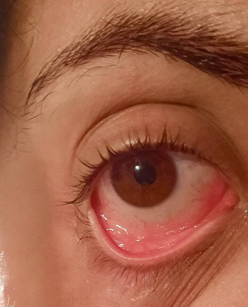
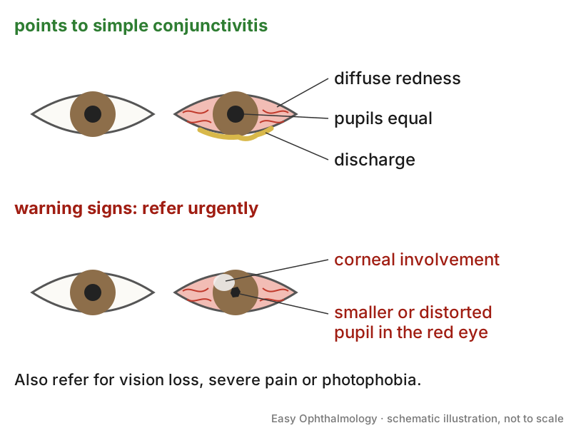
- Common causes: conjunctivitis (the most common), blepharitis, corneal abrasion, foreign body, subconjunctival haemorrhage, keratitis, iritis, glaucoma, chemical burn and scleritis [S2].
- Groups by seriousness: ocular surface disease (generally benign), intraocular disease such as uveitis, glaucoma, scleritis and trauma (ophthalmically severe), and extraocular disease such as orbital cellulitis and cavernous sinus thrombosis (systemically severe) [S11].
- Conjunctivitis: viral, bacterial or allergic; it is usually self-limiting but symptoms can last up to three weeks [S10]. Bacterial conjunctivitis was more common in children (71% vs 16% viral), and viral more common in adults (78% vs 16% bacterial), on limited evidence [S8].
Confirm it
- Basic examination: visual acuity, pupils, visual fields, penlight or slit-lamp examination of the anterior segment and direct fundoscopy [S4]. The cause is diagnosed through a detailed history and careful examination [S2].
- Bedside clues for conjunctivitis type:
- Favour viral: pharyngitis (LR+ 5.4 to 9.9), preauricular lymphadenopathy, contact with another person with red eye [S8].
- Favour bacterial: mucopurulent discharge (LR+ 2.1), otitis media [S8]; complete conjunctival redness obscuring tarsal vessels (LR 4.6), observed purulent discharge (LR 3.9) and both eyes matted in the morning (LR 3.6) [S5].
- Against bacterial: no redness visible from 20 feet (LR 0.2) and no morning gluing of either eye (LR 0.3) [S5].
- Swabs: because clinical diagnosis is difficult, a swab for PCR and culture can help confirm the cause [S10].
Manage it
Before acting on this section, check the national and local guidelines, product information and regulatory approvals that apply where you practise. See the medical disclaimer.
Tier A (primary care / no specialist)
- Screen every patient against the red flags above; red flags are especially valuable where investigations are minimal [S7].
- Simple conjunctivitis: manage conservatively. Prescribe antibiotics after a delay if symptoms do not improve within 3 days, or not at all, as recommended in [S1]. In an individual patient data meta-analysis of 3 trials (622 patients), 80% given antibiotics and 74% of controls were cured at day 7; benefit was small and seen mainly with purulent discharge or mild redness [S3].
- Give written information on the self-limiting course; this was the most effective way to manage the condition and improve satisfaction [S1].
- Contact lens wearer with suspected bacterial keratitis and no confirmed timely ophthalmology review: frequent topical fluoroquinolone may be needed, because commonly used antibiotics may not cover the cornea adequately [S12]. Doses are not given in the abstract; follow local formularies.
- Trauma and chemicals: irrigate chemical injuries immediately; cover a suspected globe injury with a metal shield and refer [S4].
Tier B (general eye clinic)
- Examine the anterior segment with the slit lamp to identify keratitis, uveitis, glaucoma or scleritis [S4] [S11].
- Treatment follows the underlying cause [S2].
Tier C (specialist centre)
- Not specifically addressed by these sources; see the condition-specific articles in this library.
Refer when
| Situation | Urgency |
|---|---|
| Red eye with vision loss, severe pain, corneal involvement, distorted pupil, trauma or recent surgery | Immediate or same-day ophthalmology referral [S2] [S6] |
| Chemical injury | Immediate irrigation, then same-day referral [S4] |
| Contact lens wearer with a painful red eye | Same-day (possible microbial keratitis) [S12] |
| Suspected orbital cellulitis or cavernous sinus thrombosis | Immediate [G1] [S11] |
| Conjunctivitis not improving after 3 days | Within days: review and consider antibiotics [S1] |
| Conjunctivitis symptoms beyond 3 weeks | Routine to an eye clinic; timing per your local service or specialist advice [S10] |
Pitfalls
- Labelling every red eye as conjunctivitis [S11].
- Skipping visual acuity and the pupil check [S4] [S5].
- Missing early keratitis in contact lens wearers [S12].
- Routine antibiotics for conjunctivitis may delay diagnosis of other serious red eye conditions [S1].
- Overconfidence: many emergency department foundation doctors lack confidence with fundoscopy and the slit lamp; formal eye training in emergency departments was considered inadequate [S9].
Evidence notes
| Source | Date | Design |
|---|---|---|
| [S1] | 2009 | Evidence review (level I and II evidence) |
| [S2] | 2010 | Narrative review (primary care) |
| [S3] | 2011 | Individual patient data meta-analysis, 3 trials |
| [S4] | 2013 | Narrative review |
| [S5] | 2015 | Systematic review of bedside diagnosis |
| [S6] | 2015 | Narrative review |
| [S7] | 2018 | Narrative review (red flags in primary care) |
| [S8] | 2022 | Systematic review and meta-analysis, 32 studies |
| [S9] | 2024 | Systematic review, 7 studies, 956 UK doctors |
| [S10] | 2024 | Narrative review |
| [S11] | 2026 | Expert review |
| [S12] | 2026 | Narrative review (contact lens users) |
- The red-flag list for referral is expert opinion from narrative reviews [S2] [S11]; only the bedside-sign likelihood ratios come from systematic reviews [S5] [S8], and some confidence intervals are very wide (photophobia to indirect light: LR 28.8, 95% CI 1.8 to 459) [S5].
- Prevalence of viral versus bacterial conjunctivitis in adults comes from a single study [S8].
- Not covered by these sources: adenoviral outbreak control, neonatal conjunctivitis, allergic conjunctivitis treatment and specific drug doses.
Related articles
References
See the numbered source list below. Each marker [S#] links to its PubMed and DOI record.
Sources
- [S1] Visscher KL, Hutnik CM, Thomas M Evidence-based treatment of acute infective conjunctivitis: Breaking the cycle of antibiotic prescribing. Canadian family physician Medecin de famille canadien. (2009). PubMed 19910590
- [S2] Cronau H, Kankanala RR, Mauger T Diagnosis and management of red eye in primary care. American family physician. (2010). PubMed 20082509
- [S3] Jefferis J, Perera R, Everitt H, van Weert H, Rietveld R, Glasziou P, et al. Acute infective conjunctivitis in primary care: who needs antibiotics? An individual patient data meta-analysis. The British journal of general practice : the journal of the Royal College of General Practitioners. (2011). PubMed 22152728 · doi:10.3399/bjgp11x593811
- [S4] Gelston CD Common eye emergencies. American family physician. (2013). PubMed 24364572
- [S5] Narayana S, McGee S Bedside Diagnosis of the 'Red Eye': A Systematic Review. The American journal of medicine. (2015). PubMed 26169885 · doi:10.1016/j.amjmed.2015.06.026
- [S6] Dunlop AL, Wells JR Approach to Red Eye for Primary Care Practitioners. Primary care. (2015). PubMed 26319338 · doi:10.1016/j.pop.2015.05.002
- [S7] Ramanayake RPJC, Basnayake BMTK Evaluation of red flags minimizes missing serious diseases in primary care. Journal of family medicine and primary care. (2018). PubMed 30090770 · doi:10.4103/jfmpc.jfmpc_510_15
- [S8] Johnson D, Liu D, Simel D Does This Patient With Acute Infectious Conjunctivitis Have a Bacterial Infection?: The Rational Clinical Examination Systematic Review. JAMA. (2022). PubMed 35699701 · doi:10.1001/jama.2022.7687
- [S9] Mendall J, Tolley A, Parisi V, Hornby S, Brown R, Nowak V Confidence of Emergency Department doctors in managing ophthalmic emergencies: a systematic review. Eye (London, England). (2024). PubMed 38729998 · doi:10.1038/s41433-024-03115-z
- [S10] Gin C, Crock C, Wells K Conjunctivitis: A review. Australian journal of general practice. (2024). PubMed 39499843 · doi:10.31128/ajgp-09-23-6960
- [S11] Arranz-Marquez E, Garcia-Gonzalez M, Teus MA Red eye: diagnostic criteria, signs of alert and therapeutic approach. Medicina clinica. (2026). PubMed 42000166 · doi:10.1016/j.medcli.2026.107433
- [S12] Ghalibafan S, Ferguson R, Ufongene C, Selvam A, Saperstein M, Sabater AL, et al. Acute red eye in contact lens users: Clinical pearls for frontline clinicians. The American journal of medicine. (2026). PubMed 42448138 · doi:10.1016/j.amjmed.2026.06.039
Guidelines
- [G1] NHS Scotland (Community Eyecare Guidelines, eyes.nhs.scot). 2.2 Cellulitis, Orbital (2026). https://www.eyes.nhs.scot/eyecare-guidelines/section-2-orbitlacrimal-system/22-cellulitis-orbital/