Educational content only. Not personalised medical advice. Clinical decisions remain the responsibility of the treating clinician.
Optic disc assessment in glaucoma
Glaucoma is diagnosed traditionally from optic nerve head damage seen on ophthalmoscopy or photography, from matching visual field damage, or both [S2]. Optic disc photographs are accessible and cheap but subjectively graded, while OCT is accurate and reproducible but costly and less portable [S8]. Glaucoma is a leading cause of irreversible blindness and is usually asymptomatic until advanced [S10].
Key points - A disc haemorrhage is associated with glaucoma progression (adjusted HR 2.03, low certainty) in a 2025 Cochrane prognostic review [S10]. - In a Cochrane review of 106 studies, OCT, HRT and GDx had similar accuracy; at about 0.95 specificity they would miss about 60 of 200 glaucoma cases among 1000 people referred from primary care [S2]. - Average RNFL thickness on OCT has moderate sensitivity (0.69 to 0.78) at high specificity (0.93 to 0.94) for manifest glaucoma [S6]. - Imaging normative databases can be flawed for atypical discs, so "red" or "green" results need critical review [S1]; myopic discs are a common source of false positives [S9]. - A swollen disc needs a different pathway: fundus photography first, then autofluorescence, ultrasound and sometimes fluorescein angiography to exclude drusen and other causes [S11].
Evidence last checked: 2026-10-17 (PubMed abstracts).
Red flags: findings that need urgent action
- Optic disc oedema. Needs further tests to exclude optic disc drusen and other causes [S11]; same-day medical or neuro-ophthalmic assessment if new or with headache or visual loss [EIC expert opinion].
- A pale or cupped disc with very high IOP or advanced field loss. Needs urgent glaucoma care to prevent further loss.
- Disc haemorrhage in a glaucoma patient or suspect. A prognostic factor for progression [S10]; bring review forward.
- Disc swelling or haemorrhage with vascular signs. OCT angiography may help in anterior ischaemic optic neuropathy and retinal vein occlusion [S11].
Recognise it: what to look for
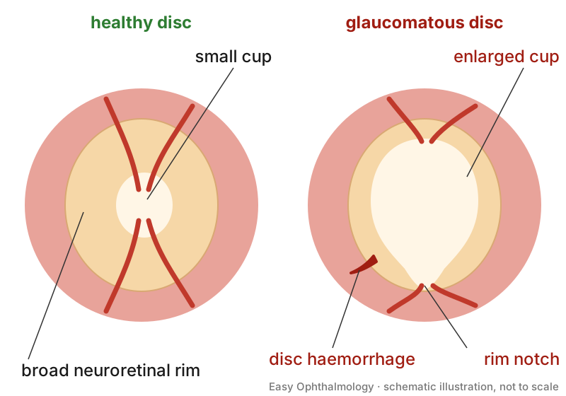
- Glaucoma is characterised by retinal nerve fibre layer loss, thinning of the neuroretinal rim and cupping of the disc, with visual field defects [S3].
- Assessment is usually required when elevated IOP or a suspicious disc is found in primary eye care [S2].
- Prognostic factors for progression: bilateral disease (moderate certainty), disc haemorrhage and female sex (low certainty); treatment was protective [S10]. Evidence for central corneal thickness, systemic hypertension and cardiovascular disease was inconsistent [S10].
- Myopia. Peripapillary atrophy and altered rim configuration in myopic eyes can resemble glaucoma [S9]. Minimum rim width, ganglion cell-inner plexiform layer thickness and the temporal raphe sign show promise in separating the two, ideally with myopia-specific normative data [S9].
Confirm it: structure and function together
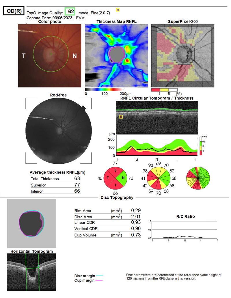
- Photography or OCT documents the disc and RNFL; imaging is an adjunct, not a replacement for clinical judgement, and artefacts occur [S1].
- In a Finnish screening sample of 3001 adults aged 45 to 49, at 97.5% specificity, sensitivity was 26% for abnormal OCT, 35% for perimetry and 44% for combined structure-function analysis; combining OCT, perimetry and defect location reached 77% sensitivity at 90% specificity [S7].
- OCT sensitivity was higher in more severe glaucoma (0.79 for mean deviation worse than -6 dB vs 0.64 for milder disease) [S2].
Manage it: assessing discs at each resource tier
Before acting on this section, check the national and local guidelines, product information and regulatory approvals that apply where you practise. See the medical disclaimer.
Tier A (primary care / no specialist)
- Fundus photography is non-invasive and low-cost [S8]. In a US telemedicine model at two health centres, 182 people had fundus and disc imaging; 34.1% had abnormal images and 18.7% unreadable images, and 82% of those referred returned for a full eye examination [S4].
- Refer any suspicious, swollen or asymmetric disc and any unreadable image (see table).
Tier B (general eye clinic)
- Assess optic nerve structure together with perimetry: early diagnosis relies on both [S3].
- Record a baseline disc photograph where possible, because subjective grading is inconsistent [S8].
- Look specifically for disc haemorrhages and note bilateral disease [S10].
- Glaucoma screening was found to be cost-effective in India and China, where most glaucoma remains undiagnosed [S5].
Tier C (specialist centre)
- Use OCT RNFL and ganglion cell measures, knowing that average RNFL thickness misses some manifest glaucoma [S6] and that accuracy in the primary studies may be overestimated by case-control designs [S2].
- Check scans for artefacts and atypical anatomy before trusting colour-coded results [S1]; use myopia-adjusted parameters in myopic eyes [S9].
- Deep learning on disc photographs or OCT appears able to diagnose glaucoma more accurately than human graders [S5]; AI trained on OCT-derived data may avoid copying human grading errors [S8].
- For swollen or atypical discs, use multimodal imaging: autofluorescence, ultrasound, OCT angiography and, when needed, fluorescein angiography [S11].
Refer when
| Situation | Urgency |
|---|---|
| New disc swelling, especially with headache or visual loss | Same-day [EIC expert opinion] |
| Glaucomatous disc with very high IOP or advanced field loss | Within days [G1] |
| Suspicious disc (large cup, rim notch, haemorrhage, asymmetry) with normal IOP [S10] | Routine; timing per your local service or specialist advice |
| Unreadable disc photograph in a screening programme [S4] | Routine, for full examination; timing per your local service or specialist advice |
Pitfalls
- Trusting colour-coded imaging printouts without looking at the scan [S1].
- Labelling myopic discs as glaucoma, or missing glaucoma in myopia [S9].
- Relying on OCT alone: it misses a meaningful share of cases [S2] [S6].
- Ignoring a small disc haemorrhage [S10].
- Missing the separate work-up a swollen disc needs [S11].
- Expecting a high positive predictive value from screening in low-prevalence populations [S7] [S8].
Evidence notes
| Source | Date | Design |
|---|---|---|
| [S1] | 2012 | Narrative review |
| [S2] | 2015 | Cochrane diagnostic accuracy review, 106 studies |
| [S3] | 2016 | Narrative review (developing countries) |
| [S4] | 2018 | Telemedicine screening study, 182 participants |
| [S5] | 2020 | Narrative review |
| [S6] | 2021 | Overview of 4 systematic reviews |
| [S7] | 2022 | Population screening study, 3001 participants |
| [S8] | 2024 | Narrative review |
| [S9] | 2025 | Narrative review |
| [S10] | 2025 | Cochrane prognostic review, 22 studies, 6082 participants |
| [S11] | 2026 | Narrative review |
- In the Cochrane accuracy review, 103 of 106 studies were case-control, which can overestimate accuracy, and 24 were manufacturer-sponsored [S2].
- In the prognostic review, 19 of 22 studies were at high risk of bias [S10].
- The screening study was in Caucasian adults aged 45 to 49 [S7], and the telemedicine study in predominantly African American participants in the US [S4]; generalisability to other settings is uncertain.
- Not covered by these sources: cup-to-disc ratio thresholds, the ISNT rule, disc size grading, direct ophthalmoscopy technique and agreement between observers.
Related articles
- Primary Open-Angle Glaucoma: Diagnosis and Treatment Targets
- Idiopathic Intracranial Hypertension and Papilloedema
- OCT interpretation: macula and retinal nerve fibre layer
References
See the numbered source list below. Each marker [S#] links to its PubMed and DOI record.
Sources
- [S1] Chong GT, Lee RK Glaucoma versus red disease: imaging and glaucoma diagnosis. Current opinion in ophthalmology. (2012). PubMed 22262083 · doi:10.1097/icu.0b013e32834ff431
- [S2] Michelessi M, Lucenteforte E, Oddone F, Brazzelli M, Parravano M, Franchi S, et al. Optic nerve head and fibre layer imaging for diagnosing glaucoma. The Cochrane database of systematic reviews. (2015). PubMed 26618332 · doi:10.1002/14651858.cd008803.pub2
- [S3] Butt NH, Ayub MH, Ali MH Challenges in the management of glaucoma in developing countries. Taiwan journal of ophthalmology. (2016). PubMed 29018725 · doi:10.1016/j.tjo.2016.01.004
- [S4] Hark L, Acito M, Adeghate J, Henderer J, Okudolo J, Malik K, et al. Philadelphia Telemedicine Glaucoma Detection and Follow-up Study: Ocular Findings at Two Health Centers. Journal of health care for the poor and underserved. (2018). PubMed 30449754 · doi:10.1353/hpu.2018.0103
- [S5] Tan NYQ, Friedman DS, Stalmans I, Ahmed IIK, Sng CCA Glaucoma screening: where are we and where do we need to go? Current opinion in ophthalmology. (2020). PubMed 31904596 · doi:10.1097/icu.0000000000000649
- [S6] Michelessi M, Li T, Miele A, Azuara-Blanco A, Qureshi R, Virgili G Accuracy of optical coherence tomography for diagnosing glaucoma: an overview of systematic reviews. The British journal of ophthalmology. (2021). PubMed 32493760 · doi:10.1136/bjophthalmol-2020-316152
- [S7] Karvonen E, Stoor K, Luodonpää M, Hägg P, Leiviskä I, Liinamaa J, et al. Combined structure-function analysis in glaucoma screening. The British journal of ophthalmology. (2022). PubMed 34230023 · doi:10.1136/bjophthalmol-2021-319178
- [S8] Beniz LAF, Campos VP, Medeiros FA Optical Coherence Tomography Versus Optic Disc Photo Assessment in Glaucoma Screening. Journal of glaucoma. (2024). PubMed 38546240 · doi:10.1097/ijg.0000000000002392
- [S9] Kim YW, Park KH How to diagnose glaucoma in myopic eyes by detecting structural changes? Asia-Pacific journal of ophthalmology (Philadelphia, Pa.). (2025). PubMed 39798605 · doi:10.1016/j.apjo.2025.100135
- [S10] Piyasena MP, Daka Q, Qureshi R, Roberti G, Michelessi M, Liu SH, et al. Prognostic factors associated with progression of open-angle glaucoma in adults. The Cochrane database of systematic reviews. (2025). PubMed 41370133 · doi:10.1002/14651858.cd015436.pub2
- [S11] Fossataro C, Savastano MC, Mottola F, De Dominicis F, Campaniello G, Cocuzza M, et al. Diagnostic Role of Multimodal Imaging in Optic Disc Diseases. Diagnostics (Basel, Switzerland). (2026). PubMed 42650994 · doi:10.3390/diagnostics16162591
Guidelines
- [G1] NHS Scotland (Community Eyecare Guidelines, eyes.nhs.scot). 6.2 Glaucoma, Primary Open Angle (n.d.). https://www.eyes.nhs.scot/eyecare-guidelines/section-6-glaucoma/62-glaucoma-primary-open-angle/