Educational content only. Not personalised medical advice. Clinical decisions remain the responsibility of the treating clinician.
Intraocular foreign body
An intraocular foreign body (IOFB) is a severe complication of ocular trauma, seen worldwide, that carries significant ocular morbidity [S10]. Most IOFBs are metallic and acquired by hammering, often at work without eye protection [S8] [S9]. A retained IOFB raises the risk of endophthalmitis, and a retained iron IOFB can cause siderosis and late vision loss [S1] [S3] [S7].
Key points - Suspect an IOFB after any hammering injury, even if the eye looks almost normal; 43% of IOFBs in one series were seen only on CT [S8]. - Non-contrast thin-cut CT is the most practical first imaging test [S5] [S11]. A negative CT does not exclude an IOFB [S12]. - MRI is dangerous when a metallic IOFB may be present [S4]. - Intravitreal antibiotics at repair reduced endophthalmitis in eyes with an IOFB in an RCT [S1]. - Same-stage IOFB removal with primary repair within 24 hours is recommended whenever feasible (expert review) [S11].
Evidence last checked: 2026-10-17 (PubMed abstracts).
Red flags
- History of hammering metal, work-related injury or no eye protection [S8] [S9].
- Signs of endophthalmitis: pain, hypopyon, vitritis or retinal periphlebitis [S3].
- Injury in a rural setting, lens disruption or delayed wound closure, which are risk factors for endophthalmitis together with a retained IOFB [S3].
- Vegetable matter or toxic metals, which change surgical approach and timing [S10].
- Planned MRI in a patient with a possible metallic IOFB: a hidden metallic IOFB caused intense ocular pain during MRI despite screening [S4].
- Late signs of a missed iron IOFB: iris heterochromia, a dilated pupil, cataract and pigmentary retinal degeneration [S7].
Recognise it
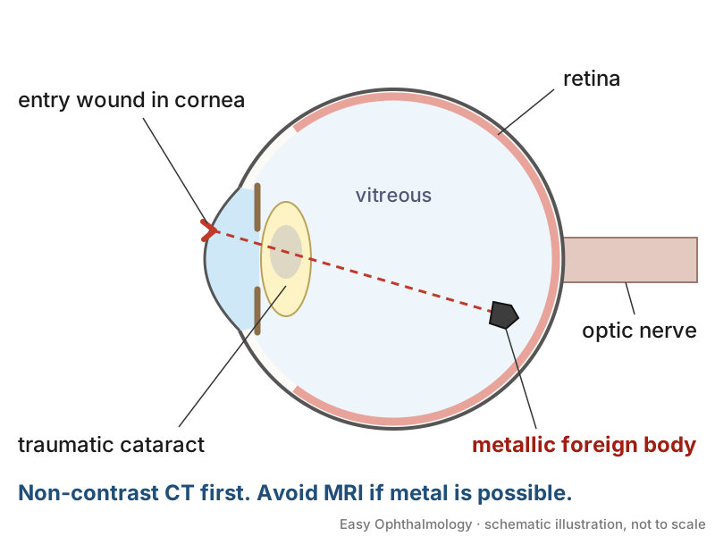
- Patients are mostly young men: 96.8% male in a Canadian series and 100% in an Irish series [S8] [S9]. In the Irish series the cornea was the route of entry in 70% [S8].
- In the Canadian series 87.5% of IOFBs were metallic, 46.9% were work-related, and traumatic cataract was the most common complication (84.4%) [S9].
- Ocular siderosis may develop from 18 days to years after injury; the most common cause is delayed presentation or a missed IOFB [S7]. ERG b-wave reduction appears before clinical signs [S7].
Confirm it
- Clinical detection is not enough: 52% of IOFBs were clinically detectable and 43% were identifiable only on CT [S8].
- CT: the most practical first step because it detects a wide range of IOFB types at small sizes [S5]. Non-contrast helical CT gives information on size, shape and location [S2]. In a systematic review CT was 69% sensitive for IOFB against intraoperative findings, so a negative CT does not exclude an IOFB [S12]. In a small Canadian series CT detected 100% of IOFBs and B-scan 87.5% [S9].
- Ultrasound: B-scan was 86% sensitive for IOFB compared with CT, but its safety with respect to pressure on an open globe was not studied [S12]. In a meta-analysis, emergency-practitioner point-of-care ultrasound showed high sensitivity and specificity for IOFB, but few studies assessed IOFB and more study is needed [S6].
- MRI and ultrasound are adjuncts, useful for wood, plastic and glass IOFBs [S5]; avoid MRI until a metallic IOFB has been excluded [S4].
- Assess for concomitant life-threatening injuries first [S2].
Manage it
Before acting on this section, check the national and local guidelines, product information and regulatory approvals that apply where you practise. See the medical disclaimer.
Tier A (primary care / no specialist)
- Treat any suspected IOFB as an open-globe injury: protect the eye without pressure and arrange immediate transfer to an eye surgeon.
- Do not request MRI [S4].
- Record the mechanism (such as hammering) and the material if known; composition affects surgical planning [S10].
Tier B (general eye clinic)
- Obtain CT where available [S5] [S11].
- Antibiotic prophylaxis. Perioperative systemic and intravitreal antibiotics may reduce endophthalmitis (expert review) [S11]. Perioperative systemic and topical third- or fourth-generation fluoroquinolones may play a role (expert review) [S2]. Doses are not given in these abstracts; follow local formularies.
- Transfer to a vitreoretinal service without delay [S11].
Tier C (specialist centre)
- Timing. Same-stage removal with primary repair within 24 hours is recommended whenever feasible [S11]. Delayed versus immediate removal depends on the patient's medical status, the operating facilities and staff available, and whether endophthalmitis is present [S2]. The advantages and disadvantages of each approach are debated [S10].
- Intraocular antibiotics. In a multicentre RCT, endophthalmitis developed in 7 of 25 control eyes with an IOFB and in none of 27 eyes given intraocular gentamicin 40 µg and clindamycin 45 µg, as reported in [S1]. Intravitreal injection was superior to intracameral [S1]. Intravitreal antibiotics are also considered with a positive Gram stain, suspected endophthalmitis or high-risk settings [S2].
- Established endophthalmitis: intravitreal vancomycin and ceftazidime are routinely used; doses are not given in the abstract [S3].
- Surgery: posterior segment IOFBs are usually removed by pars plana vitrectomy; anterior segment IOFBs may be removed with an intraocular magnet or forceps through a limbal paracentesis [S7] [S10]. Material and size guide the instrument and extraction site [S2].
- Siderosis: remove the IOFB even after siderosis develops, because acuity and clinical signs may improve [S7].
- Follow-up: monitor closely for endophthalmitis, retinal detachment and proliferative vitreoretinopathy [S2]. In one series, 3 of 4 eyes with secondary glaucoma had retained or delayed extractions, so extended follow-up is advised [S9].
Refer when
| Situation | Urgency |
|---|---|
| Suspected or confirmed IOFB | Immediate transfer to an eye surgeon; aim for repair and removal within 24 hours [S11] |
| Pain, hypopyon or vitritis after an IOFB injury | Immediate, same-day (possible endophthalmitis) [S3] |
| Iris heterochromia or a dilated pupil after an old metal injury | Urgent, (possible siderosis); timing per your local service or specialist advice |
| Retained IOFB not removed | Routine long-term follow-up with a retina service, at intervals set by the surgeon [S9] |
Pitfalls
- Relying on a normal-looking eye or a negative CT to exclude an IOFB [S8] [S12].
- Sending a patient with a possible metallic IOFB for MRI [S4].
- Delaying repair; delayed wound closure is a risk factor for endophthalmitis [S3].
- Missing siderosis years after an injury that was never investigated [S7].
- Stopping follow-up early in retained or delayed-extraction cases [S9].
Evidence notes
| Source | Date | Design |
|---|---|---|
| [S1] | 2007 | Multicentre double-masked RCT, 346 eyes |
| [S2] | 2008 | Narrative review with management algorithm |
| [S3] | 2012 | Narrative review (post-traumatic endophthalmitis) |
| [S4] | 2018 | Case report with review |
| [S5] | 2019 | Systematic review (imaging) |
| [S6] | 2020 | Diagnostic meta-analysis, 9 studies |
| [S7] | 2021 | Literature review (siderosis) |
| [S8] | 2021 | Retrospective series, 23 eyes, Ireland |
| [S9] | 2025 | Retrospective series, 32 eyes, Canada |
| [S10] | 2024 | Narrative review |
| [S11] | 2025 | Narrative review |
| [S12] | 2026 | Systematic review, 11 diagnostic studies |
- Apart from one RCT [S1] and two diagnostic syntheses [S6] [S12], the evidence is narrative review and small retrospective series from high-income centres; management recommendations are expert opinion [S2] [S10] [S11].
- Imaging sources conflict: CT detected all IOFBs in one small series [S9] but was 69% sensitive in a systematic review [S12].
- The POCUS meta-analysis had wide confidence intervals for IOFB and excluded children [S6].
- Not covered by these sources: systemic antibiotic choice and doses, tetanus prophylaxis, plain X-ray where CT is unavailable, and outcomes in low-resource settings.
Related articles
References
See the numbered source list below. Each marker [S#] links to its PubMed and DOI record.
Sources
- [S1] Soheilian M, Rafati N, Mohebbi MR, Yazdani S, Habibabadi HF, Feghhi M, et al. Prophylaxis of acute posttraumatic bacterial endophthalmitis: a multicenter, randomized clinical trial of intraocular antibiotic injection, report 2. Archives of ophthalmology (Chicago, Ill. : 1960). (2007). PubMed 17420365 · doi:10.1001/archopht.125.4.460
- [S2] Yeh S, Colyer MH, Weichel ED Current trends in the management of intraocular foreign bodies. Current opinion in ophthalmology. (2008). PubMed 18408498 · doi:10.1097/icu.0b013e3282fa75f1
- [S3] Ahmed Y, Schimel AM, Pathengay A, Colyer MH, Flynn HW Endophthalmitis following open-globe injuries. Eye (London, England). (2012). PubMed 22134598 · doi:10.1038/eye.2011.313
- [S4] Mamas N, Andreanos K, Brouzas D, Diagourtas A, Droulias A, Georgalas I, et al. Acute ocular pain during magnetic resonance imaging due to retained intraocular metallic foreign body: the role of ultrasonography and ultrasound biomicroscopy in diagnosis and management of this condition. Journal of ultrasound. (2018). PubMed 29500812 · doi:10.1007/s40477-018-0289-2
- [S5] Rong AJ, Fan KC, Golshani B, Bobinski M, McGahan JP, Eliott D, et al. Multimodal imaging features of intraocular foreign bodies. Seminars in ophthalmology. (2019). PubMed 31609153 · doi:10.1080/08820538.2019.1674894
- [S6] Propst SL, Kirschner JM, Strachan CC, Roumpf SK, Menard LM, Sarmiento EJ, et al. Ocular Point-of-Care Ultrasonography to Diagnose Posterior Chamber Abnormalities: A Systematic Review and Meta-analysis. JAMA network open. (2020). PubMed 32074291 · doi:10.1001/jamanetworkopen.2019.21460
- [S7] Casini G, Sartini F, Loiudice P, Benini G, Menchini M Ocular siderosis: a misdiagnosed cause of visual loss due to ferrous intraocular foreign bodies-epidemiology, pathogenesis, clinical signs, imaging and available treatment options. Documenta ophthalmologica. Advances in ophthalmology. (2021). PubMed 32949328 · doi:10.1007/s10633-020-09792-x
- [S8] Bourke L, Bourke E, Cullinane A, O'Connell E, Idrees Z Clinical outcomes and epidemiology of intraocular foreign body injuries in Cork University Hospital, Ireland: an 11-year review. Irish journal of medical science. (2021). PubMed 33230610 · doi:10.1007/s11845-020-02443-9
- [S9] Al-Ani A, Bondok MS, Madjedi K, Kherani S, Kherani A Clinical outcomes and characterization of intraocular foreign body injuries from a Canadian centre: a 20-year retrospective study and literature review. Canadian journal of ophthalmology. Journal canadien d'ophtalmologie. (2025). PubMed 39095034 · doi:10.1016/j.jcjo.2024.05.019
- [S10] Ohlhausen M, Menke BA, Begley J, Kim S, Debiec MR, Conrady CD, et al. Advances in the management of intraocular foreign bodies. Frontiers in ophthalmology. (2024). PubMed 39285858 · doi:10.3389/fopht.2024.1422466
- [S11] Au ACK, Wong DHT, Li KKW Updates on the clinical management of intraocular foreign body (IOFB). Asia-Pacific journal of ophthalmology (Philadelphia, Pa.). (2025). PubMed 40398511 · doi:10.1016/j.apjo.2025.100208
- [S12] Mair J, Bush L, Halliday S, McMaster D, Sellon E, Colyer MH, et al. Use of preoperative imaging in open globe injury management: a systematic review. The British journal of ophthalmology. (2026). PubMed 41423323 · doi:10.1136/bjo-2025-327387