Educational content only. Not personalised medical advice. Clinical decisions remain the responsibility of the treating clinician.
Gonioscopy and anterior-segment imaging of the angle
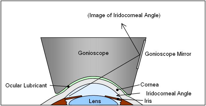
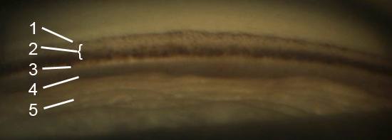
Slit-lamp gonioscopy is the reference standard for evaluating the anterior chamber angle and is essential for diagnosing angle closure disease [S6]. Its weaknesses are a steep learning curve, poor agreement between examiners and poor documentation [S4] [S6]. Anterior segment OCT (AS-OCT), ultrasound biomicroscopy (UBM) and goniophotography add objective and reproducible information but, in an American Academy of Ophthalmology assessment, none was a substitute for gonioscopy [S1].
Key points - Gonioscopy remains the primary method of angle assessment; imaging is an adjunct with better reproducibility and documentation [S2]. - Deep learning applied to AS-OCT detected angle closure with pooled sensitivity of 94.0% and specificity of 93.6% against gonioscopy (6 studies, 5269 patients) [S9]. - Where gonioscopy and AS-OCT are unavailable, anterior chamber depth estimated by the oblique flashlight test or van Herick technique is a screening option [S7]. - Only UBM images structures behind the iris and it can be used when an opaque cornea prevents gonioscopy [S1] [S5]. - In the Zhongshan Angle-Closure Prevention (ZAP) trial, laser iridotomy cut progression from suspect to primary angle closure by about two thirds over 14 years, but absolute risk was low [S8].
Evidence last checked: 2026-10-17 (PubMed abstracts).
Red flags: findings that need urgent action
- Closed angle with high IOP, pain, corneal oedema or a mid-dilated pupil. Possible acute angle closure: same-day treatment [G1].
- Peripheral anterior synechiae or IOP above 24 mm Hg in an angle closure suspect. These define progression to primary angle closure in the ZAP trial [S8] [S11].
- Iridotrabecular contact (ITC) in the nasal sector on AS-OCT. Associated with a hazard ratio of 4.68 for progression to angle closure over 72 months [S11].
- Angle not visible because of corneal opacity. Use UBM to define the mechanism [S5].
Recognise it: who needs angle assessment
- Angle assessment is intrinsic to glaucoma diagnosis and treatment and has a role in prevention [S4].
- Primary angle closure glaucoma is a leading cause of irreversible blindness, particularly in Asia [S7].
- In ZAP control eyes, higher IOP, shallower limbal anterior chamber depth and greater central anterior chamber depth were associated with progression [S8].
Confirm it: methods compared
| Method | What it adds | Limits |
|---|---|---|
| Slit-lamp gonioscopy | Reference standard; qualitative view [S6] | Learning curve, examiner disagreement, poor documentation [S6] |
| Automated gonioscopy (GS-1, NGS-1) | Real-colour circumferential photographs; telemedicine and deep learning potential [S6] [S7] | NGS-1 is the only one with dedicated angle software [S7] |
| AS-OCT | Non-contact, quantitative; imaging in darkness detects appositional closure more sensitively [S1] | Not a substitute for gonioscopy [S1] |
| UBM | Only method imaging behind the iris; works through opaque cornea [S1] [S5] | Contact method, less suited to mass screening [S1] |
| Scheimpflug, scanning peripheral ACD analyser | Non-contact ACD measurement, suits screening [S1] [S7] | Indirect: does not image the angle itself [S1] |
| Van Herick, oblique flashlight | Simple estimate of anterior chamber depth [S7] | Screening only [S7] |
- An automated algorithm grading trabecular pigmentation on 360° gonioscopy images agreed substantially with manual grading (kappa 0.70) in 75 open-angle eyes; pigment was densest inferiorly [S3].
- An international consensus (APOSTEL-AS, 2025) agreed 31 standard terms for anatomical landmarks on AS-OCT to improve reporting [S10].
Manage it: doing and acting on the test at each tier
Before acting on this section, check the national and local guidelines, product information and regulatory approvals that apply where you practise. See the medical disclaimer.
Tier A (primary care / no specialist)
- Without a slit lamp or gonioscope, use the oblique flashlight test as a screen for a shallow anterior chamber [S7].
- Refer people with a suspected shallow chamber for gonioscopy (see table).
Tier B (general eye clinic)
- Estimate limbal anterior chamber depth with the van Herick technique [S7], and perform gonioscopy, the reference standard, whenever angle closure is suspected [S6].
- Record findings in a structured way, because poor documentation is a known weakness of gonioscopy [S6].
- Where AS-OCT with deep learning is available, it may support screening in populations without access to experienced gonioscopists [S9].
Tier C (specialist centre)
- Use AS-OCT or UBM as adjuncts to gonioscopy for mechanism, risk assessment and documentation [S2]; UBM for opaque corneas and to image structures behind the iris [S1] [S5].
- Look for nasal or superior ITC on AS-OCT: ITC length was associated with progression (HR 1.64 per 250 µm nasally), whereas cumulative gonioscopy score was not [S11].
- For angle closure suspects, weigh the two-thirds relative reduction in progression after iridotomy against the low cumulative risk in community-based suspects [S8]. Iridotomy-treated eyes had more severe nuclear cataract at 14 years [S8].
- Use standard AS-OCT nomenclature in reports and research [S10].
Refer when
| Situation | Urgency |
|---|---|
| Suspected acute angle closure (closed angle, high IOP, pain, oedema) | Immediate, same-day [G1] |
| Shallow chamber on flashlight or van Herick test, no symptoms [S7] | Routine, for gonioscopy; timing per your local service or specialist advice |
| Angle closure suspect with peripheral anterior synechiae or raised IOP [S8] | Within a week, to a glaucoma service [G2] |
| Angle not assessable (opaque cornea) with raised IOP [S5] | Urgent to a centre with UBM; timing per your local service or specialist advice |
Pitfalls
- Treating an imaging result as a replacement for gonioscopy [S1].
- Forgetting that lighting matters: OCT imaging in complete darkness detects appositional closure more sensitively [S1].
- Relying on van Herick alone to diagnose angle closure [S7].
- Leaving gonioscopy findings undocumented [S6].
- Assuming every angle closure suspect will progress: the 14-year risk was low [S8].
Evidence notes
| Source | Date | Design |
|---|---|---|
| [S1] | 2013 | AAO Ophthalmic Technology Assessment, 79 studies |
| [S2] | 2018 | Narrative review and perspective |
| [S3] | 2020 | Clinical study, 75 eyes |
| [S4] | 2020 | Narrative review |
| [S5] | 2021 | Narrative review |
| [S6] | 2021 | Narrative review (single device focus) |
| [S7] | 2022 | Narrative review |
| [S8] | 2023 | RCT extended follow-up, 889 patients, 14 years |
| [S9] | 2024 | Systematic review and meta-analysis, 6 studies |
| [S10] | 2025 | Expert consensus, 14 panellists |
| [S11] | 2026 | Retrospective cohort within an RCT, 825 eyes |
- ZAP lost 390 treated and 388 control eyes to follow-up over 14 years [S8], and both ZAP analyses are in Chinese participants aged 50 to 70 years [S8] [S11].
- The AAO assessment predates deep learning studies and widely available swept-source AS-OCT [S1]; the meta-analysis included only 6 studies [S9].
- Sources differ in emphasis: one review argues AS-imaging could become first-line screening [S2]; the AAO assessment found no method a substitute for gonioscopy [S1].
- A Brazilian guideline on angle-closure diagnosis appeared in the search but had no abstract and was not used.
- Not covered by these sources: step-by-step technique for indentation gonioscopy, lens choice, angle grading systems (Shaffer, Spaeth), and van Herick grading cut-offs.
Related articles
References
See the numbered source list below. Each marker [S#] links to its PubMed and DOI record.
Sources
- [S1] Smith SD, Singh K, Lin SC, Chen PP, Chen TC, Francis BA, et al. Evaluation of the anterior chamber angle in glaucoma: a report by the american academy of ophthalmology. Ophthalmology. (2013). PubMed 23978623 · doi:10.1016/j.ophtha.2013.05.034
- [S2] Chansangpetch S, Rojanapongpun P, Lin SC Anterior Segment Imaging for Angle Closure. American journal of ophthalmology. (2018). PubMed 29352976 · doi:10.1016/j.ajo.2018.01.006
- [S3] Matsuo M, Pajaro S, De Giusti A, Tanito M Automated anterior chamber angle pigmentation analyses using 360° gonioscopy. The British journal of ophthalmology. (2020). PubMed 31471328 · doi:10.1136/bjophthalmol-2019-314320
- [S4] Riva I, Micheletti E, Oddone F, Bruttini C, Montescani S, De Angelis G, et al. Anterior Chamber Angle Assessment Techniques: A Review. Journal of clinical medicine. (2020). PubMed 33255754 · doi:10.3390/jcm9123814
- [S5] Potop V, Coviltir V, Schmitzer S, Dragosloveanu CDM, Ionescu CI, Burcel MG, et al. Ultrasound biomicroscopy in glaucoma assessment. Romanian journal of ophthalmology. (2021). PubMed 34179574 · doi:10.22336/rjo.2021.24
- [S6] Cutolo CA, Bonzano C, Scotto R, Iester M, Bagnis A, Pizzorno C, et al. Moving beyond the Slit-Lamp Gonioscopy: Challenges and Future Opportunities. Diagnostics (Basel, Switzerland). (2021). PubMed 34943516 · doi:10.3390/diagnostics11122279
- [S7] Porporato N, Bell KC, Perera SA, Aung T Non-optical coherence tomography modalities for assessment of angle closure. Taiwan journal of ophthalmology. (2022). PubMed 36660111 · doi:10.4103/tjo.tjo_41_21
- [S8] Yuan Y, Wang W, Xiong R, Zhang J, Li C, Yang S, et al. Fourteen-Year Outcome of Angle-Closure Prevention with Laser Iridotomy in the Zhongshan Angle-Closure Prevention Study: Extended Follow-up of a Randomized Controlled Trial. Ophthalmology. (2023). PubMed 37030454 · doi:10.1016/j.ophtha.2023.03.024
- [S9] Olyntho MAC, Jorge CAC, Castanha EB, Gonçalves AN, Silva BL, Nogueira BV, et al. Artificial Intelligence in Anterior Chamber Evaluation: A Systematic Review and Meta-Analysis. Journal of glaucoma. (2024). PubMed 38747721 · doi:10.1097/ijg.0000000000002428
- [S10] Fraser AS, Ang M, Bellchambers A, Chu CJ, Denniston AK, Downie LE, et al. Proposed Nomenclature for Landmarks in Anterior-Segment OCT: The APOSTEL-AS Panel Consensus. JAMA ophthalmology. (2025). PubMed 40810988 · doi:10.1001/jamaophthalmol.2025.2414
- [S11] Hong KL, Xu BY, Wang W, Cho A, Foster PJ, Jiang Y, et al. Iris Trabecular Contact on OCT in Angle Closure Progression. JAMA ophthalmology. (2026). PubMed 41642600 · doi:10.1001/jamaophthalmol.2025.6067
Guidelines
- [G1] NHS Scotland Community Eyecare Guidelines. 6.4 Glaucoma, Acute Angle Closure (n.d.). https://www.eyes.nhs.scot/eyecare-guidelines/section-6-glaucoma/64-glaucoma-acute-angle-closure/
- [G2] NHS Scotland Community Eyecare Guidelines. 6.3 Glaucoma, Primary Angle Closure (n.d.). https://www.eyes.nhs.scot/eyecare-guidelines/section-6-glaucoma/63-glaucoma-primary-angle-closure/