Educational content only. Not personalised medical advice. Clinical decisions remain the responsibility of the treating clinician.
Eyelid and Canalicular Lacerations
Eyelid trauma ranges from simple lacerations to severe, vision-threatening injuries, and any lid injury is cosmetically sensitive [S6]. Lacrimal drainage damage is particularly hard to detect and repair [S6]. In a Nigerian tertiary hospital, eyelid laceration was the commonest oculoplastic disorder and eyelid repair the commonest oculoplastic operation [S9].
Key points - Look for occult serious injury, including the globe and orbit, in every periorbital laceration [S6]. - Restoring normal anatomical relationships of the lids and periocular structures is essential for function and appearance [S7]. - Repair monocanalicular lacerations in all cases, whether upper or lower canaliculus [S4]. - Monocanalicular stents give anatomical success of 68% to 100% (level III evidence only) [S5].
Evidence last checked: 2026-10-18 (PubMed abstracts).
Red flags
- Possible open globe or orbital fracture under a lid wound: in the Beirut blast series, eyelid lacerations (41.6% of eyes) occurred alongside orbital fractures (29.2%) and open globe injuries (20.8%) [S8]. A pooled review reported orbital fracture in 18% to 25% and globe trauma in 10% to 15% of eyelid trauma [S11].
- Medial lid laceration: suspect canalicular injury, which is easily missed [S6].
- Blast or shrapnel injury: many injuries presented late because services were overwhelmed [S8].
Recognise it
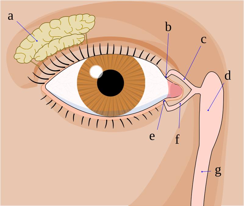
- Laceration is the most frequent form of eyelid trauma; canalicular injury and lid avulsion are less common [S11].
- Trauma was the commonest cause of oculoplastic disease in a Nigerian series, with a median patient age of 28 years [S9].
- In blasts, injuries are mostly from debris and shattered glass [S8].
Confirm it
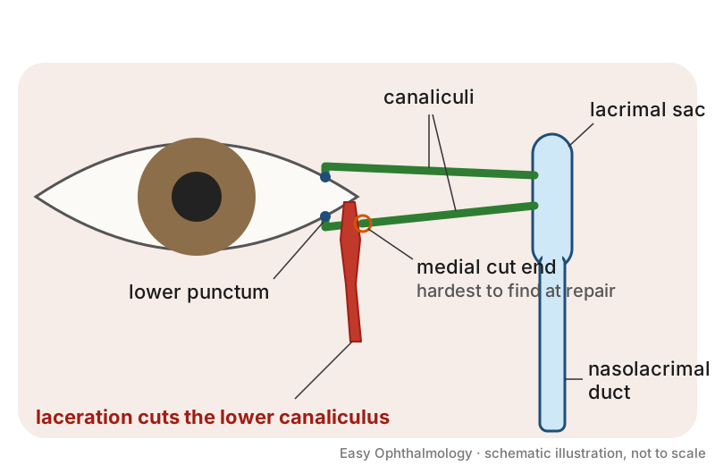
- Examine for signs of more serious, potentially occult trauma before planning lid repair [S6].
- Assess the lacrimal drainage system in any medial injury [S6].
- At repair, locating the medial cut end of the canaliculus is the hardest step, especially with medial laceration, oedema, bleeding or a narrow canaliculus [S3]. Techniques include a pigtail probe (modified probe primary success 93.48%, final 97.83%) [S1] and air injection through the intact canaliculus with the medial canthus under saline (successful in 19 cases) [S3].
Manage it
Before acting on this section, check the national and local guidelines, product information and regulatory approvals that apply where you practise. See the medical disclaimer.
Tier A (primary care / no specialist)
- Use resource-adapted guidance for ocular trauma: the EXTRACCT guideline is written for frontline workers without specialist expertise or equipment and includes dosing of ophthalmic drugs [S10].
- Do not close a lid wound before checking the eye for open globe; refer.
Tier B (general eye clinic)
- Primary layered repair is the commonest treatment [S11]; restore anatomical relationships [S7].
- In a pooled review, good-to-excellent outcomes were reported in 75% to 90% of patients repaired within 24 to 48 hours, and complications were lower with prompt treatment (5% to 10% vs 20% to 30% with delay) [S11]. This review is of uncertain quality.
- Commonest complications are epiphora, ectropion, scarring, infection and lid malposition [S11].
Tier C (specialist centre)
- Canalicular repair: reconstruct all monocanalicular lacerations [S4]. Monocanalicular stents had functional success of 79% to 100% and extrusion rates of 0% to 29% in 15 level III studies; other complications include tube displacement, granuloma, ectropion, slit punctum, fistula and infection [S5]. The optimal time for stent removal and the comparison with bicanalicular stents are not established [S5].
- Timing and logistics: in a small Chinese RCT (48 eyes), a two-step approach (finding the medial end and stenting at the slit lamp, then anastomosis in theatre) shortened time to treatment (4.3 vs 18.8 hours) and found the medial end faster, with similar 12-month patency (91.3% vs 80%) [S2].
- In the pigtail probe RCT, silicone stents were left for 4 to 6 months [S1].
Refer when
| Situation | Urgency |
|---|---|
| Lid laceration with suspected open globe | Immediate |
| Canalicular laceration or full-thickness lid margin laceration | Urgent, within 24 to 48 hours, for repair [S11] |
| Laceration with possible orbital fracture | Urgent [S8]; timing per your local service or specialist advice |
Pitfalls
- Missing a canalicular injury in a medial wound [S6].
- Leaving a lower canalicular laceration unrepaired: repair is recommended for upper and lower injuries [S4].
- Focusing on the lid and missing globe or orbital injury [S6] [S8].
- Delaying repair: delayed treatment was associated with more complications [S11].
Evidence notes
| Source | Date | Design |
|---|---|---|
| [S1] | 2009 | RCT, 87 patients, China |
| [S2] | 2013 | RCT, 48 eyes, China |
| [S3] | 2013 | Technique report, 19 cases |
| [S4] | 2016 | Narrative review |
| [S5] | 2019 | AAO evidence report, 15 level III studies |
| [S6] | 2021 | Narrative review |
| [S7] | 2021 | Narrative review |
| [S8] | 2021 | Retrospective series, Beirut blast |
| [S9] | 2022 | Retrospective hospital review, Nigeria |
| [S10] | 2026 | Expert consensus guideline, low-resource and conflict settings |
| [S11] | 2026 | Systematic review (Cureus), methods unclear |
- Thin evidence: two small single-centre RCTs on canalicular technique [S1] [S2]; everything else is level III or review.
- Check [S11]: its pooled percentages are reported as ranges derived from counts of studies, not patients, and should be verified against the full text.
- Not covered by these sources: tetanus and rabies prophylaxis, antibiotic choice, suture technique, and bite injuries.
Related articles
References
See the numbered source list below. Each marker [S#] links to its PubMed and DOI record.
Sources
- [S1] Liang T, Zhao GQ, Li YL, Yang SS, Zhang LY, Wu Y Efficiency and therapeutic effect of modified pigtail probe in anastomosing lacerated lacrimal canaliculus. Chinese journal of traumatology = Zhonghua chuang shang za zhi. (2009). PubMed 19321052
- [S2] Bi Y, Sui G, Zhou Q, Heindl LM, Bock F, Sun X, et al. Two-step retrograde closed stenting: a novel method for treating canalicular lacerations in Chinese patients. Eye (London, England). (2013). PubMed 23970028 · doi:10.1038/eye.2013.169
- [S3] Liu B, Li Y, Long C, Wang Z, Liang X, Ge J, et al. Novel air-injection technique to locate the medial cut end of lacerated canaliculus. The British journal of ophthalmology. (2013). PubMed 24037604 · doi:10.1136/bjophthalmol-2013-303822
- [S4] Kalin-Hajdu E, Cadet N, Boulos PR Controversies of the lacrimal system. Survey of ophthalmology. (2016). PubMed 26700821 · doi:10.1016/j.survophthal.2015.12.003
- [S5] Wladis EJ, Aakalu VK, Tao JP, Sobel RK, Freitag SK, Foster JA, et al. Monocanalicular Stents in Eyelid Lacerations: A Report by the American Academy of Ophthalmology. Ophthalmology. (2019). PubMed 30953742 · doi:10.1016/j.ophtha.2019.03.045
- [S6] Seol Y, Dweck M Periorbital Soft Tissue Trauma. Facial plastic surgery : FPS. (2021). PubMed 33657628 · doi:10.1055/s-0041-1725131
- [S7] Ko AC, Satterfield KR, Korn BS, Kikkawa DO Eyelid and Periorbital Soft Tissue Trauma. Oral and maxillofacial surgery clinics of North America. (2021). PubMed 34210399 · doi:10.1016/j.coms.2021.04.004
- [S8] Kheir WJ, Awwad ST, Bou Ghannam A, Khalil AA, Ibrahim P, Rachid E, et al. Ophthalmic Injuries After the Port of Beirut Blast-One of Largest Nonnuclear Explosions in History. JAMA ophthalmology. (2021). PubMed 34351374 · doi:10.1001/jamaophthalmol.2021.2742
- [S9] Adewara BA, Badmus SA, Awe OO, Onakpoya OH, Adegbehingbe BO, Adeoye AO Epidemiology and Management of Oculoplastic Disorders at a Tertiary Hospital: A 4-Year Review. West African journal of medicine. (2022). PubMed 35752971
- [S10] Butterfield E, Bolt A, Clare G, Mattia J, Tin-U AM, Ndyabawe I, et al. Explosive Weapons Trauma Care Collective (EXTRACCT) Blast Injury Clinical Practice Guideline: Ocular Trauma. World journal of surgery. (2026). PubMed 41454569 · doi:10.1002/wjs.70204
- [S11] Yadav NK, Husain A, Joshi D, Abbasi S, Arya AK, Priya P, et al. Advances in Eyelid Trauma Management: A Systematic Review of Surgical Strategies, Reconstructive Innovations, and Clinical Outcomes From 2000 to 2026. Cureus. (2026). PubMed 42375876 · doi:10.7759/cureus.109910