Educational content only. Not personalised medical advice. Clinical decisions remain the responsibility of the treating clinician.
Choroidal naevus versus small melanoma
Choroidal naevus is a common benign melanocytic tumour, usually found incidentally, with a small risk (<1%) of malignant transformation [S1]. The clinical challenge is to separate it from small choroidal melanoma, with which it overlaps as "indeterminate" lesions [S7]. Imaging-based risk factors identify the minority that need specialist review [S2].
Key points - Postequatorial choroidal naevi are present in approximately 5% of US adults aged 40 years or more [S1]. - Risk factors for transformation are summarised by the mnemonic TFSOM-DIM: thickness >2 mm, subretinal fluid, symptoms of vision loss, orange pigment, ultrasonographic hollowness and diameter >5 mm [S2]. - Five-year risk of growth into melanoma rose from 1.1% with no risk factor to 55% with five risk factors in a cohort of 3806 naevi [S2]. - Lesions with three or more risk factors should be evaluated at an experienced ocular oncology centre [S1]. - Most indeterminate lesions that grow do so in the first 2 years of observation [S8].
Evidence last checked: 2026-10-17 (PubMed abstracts).
Red flags
- Multiple risk factors. Three or more risk factors imply more than 50% chance of transformation to melanoma within 5 years in one review [S1]; in the imaging cohort, 5-year growth was 34% with three factors and 51% with four [S2].
- Documented growth. Among 8606 indeterminate tumours followed, 478 (5.6%) grew over 5 years; early growth may represent melanoma in evolution, and late growth after stability may represent malignant transformation [S8].
- Giant naevus. Basal dimension of 10 mm or more carries greater risk of transformation [S1].
- Symptoms. Flashes, floaters or blurred vision are listed risk factors [S1].
Recognise it
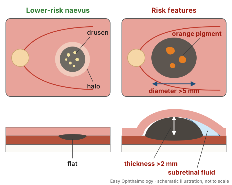
- A naevus is generally well circumscribed and pigmented; the mean basal dimension in population studies is approximately 1.25 mm [S1].
- Older risk factors also include margin less than 3 mm from the optic disc, and absence of halo and of drusen [S1].
- Many benign pigmented fundus lesions mimic naevus or melanoma, including congenital hypertrophy of the retinal pigment epithelium, RPE hyperplasia, melanocytoma and others [S5].
Confirm it
- Ultrasonography: thickness >2 mm and acoustic hollowness are risk factors [S2].
- OCT: detects subretinal fluid [S2]; enhanced-depth and swept-source OCT can show subretinal fluid, RPE disruption and dome-shaped elevation, and OCT angiography helps separate melanoma from naevi [S9]. OCT angiography showed naevi with minimal overlying macular microvascular change compared with melanoma [S1].
- Fundus autofluorescence: identifies orange pigment (lipofuscin) [S2] [S4].
- Fundus photography: measures diameter >5 mm [S2] and documents change over time; a white paper describes multimodal imaging and a possible future role for artificial intelligence in detecting transformation [S3].
- Biopsy: can be used for diagnosis of indeterminate lesions or prognosis in small melanoma [S7].
Manage it
Before acting on this section, check the national and local guidelines, product information and regulatory approvals that apply where you practise. See the medical disclaimer.
Tier A (primary care / no specialist)
- A pigmented fundus lesion seen on fundoscopy or retinal photography should be photographed if possible and assessed by an eye clinic; dilated examination and imaging are needed to grade risk [S2] [S5].
- Ask about flashes, floaters or blurred vision [S1].
Tier B (general eye clinic)
- Record diameter and margin-to-disc distance with fundus photography and look for orange pigment, fluid, drusen and halo [S1] [S2].
- Use ultrasonography and OCT where available to measure thickness and detect subretinal fluid [S2].
- Observation is reasonable for asymptomatic benign lesions [S5].
- Refer lesions with three or more risk factors, documented growth or symptoms (see Refer when) [S1] [S8].
Tier C (specialist centre)
- Indeterminate lesions can be closely monitored for growth with multimodal imaging [S7]. Treating an indeterminate lesion is weighed against visual morbidity from local treatment versus rising metastatic risk with size [S7].
- Small choroidal melanoma treated with plaque radiotherapy had tumour recurrence of 7% and 11% at 5 and 10 years and loss of 3 or more Snellen lines in 39% and 49% [S2].
- Delay matters less for small melanomas than larger ones: modelling estimates a 0.3 percentage point increase in 10-year metastatic death per month untreated for small melanomas, against 1.8 and 4.0 for medium and large tumours [S6]. Small melanomas had a mean volume doubling time of 717 days, growing naevi 6392 days [S6].
Refer when
| Situation | Urgency |
|---|---|
| Pigmented lesion with three or more risk factors, documented growth, subretinal fluid or orange pigment | Urgent to ocular oncology; timing per your local service or specialist advice [S1] |
| Lesion with one or two risk factors | Routine, for specialist imaging review; timing per your local service or specialist advice |
Pitfalls
- Relying on fundoscopy alone: several diagnostic features can be missed without multimodal imaging [S5].
- Assuming a lesion stable for years cannot change: late growth may mean malignant transformation [S8].
- Treating every indeterminate lesion: treatment carries visual morbidity [S7].
- Ignoring symptoms such as flashes or blurred vision [S1].
Evidence notes
| Source | Date | Design |
|---|---|---|
| [S1] | 2017 | Narrative review |
| [S2] | 2019 | Narrative review summarising a cohort of 3806 naevi |
| [S3] | 2021 | White paper (consensus statement) |
| [S4] | 2022 | Narrative review of autofluorescence |
| [S5] | 2024 | Narrative review of multimodal imaging |
| [S6] | 2025 | Systematic review and meta-analysis, 5 studies |
| [S7] | 2025 | Narrative review |
| [S8] | 2025 | Review of 9 largely retrospective studies |
| [S9] | 2025 | Narrative review of OCT and OCT angiography |
- Thin evidence: apart from one meta-analysis [S6], the sources are narrative reviews; risk factor figures come from single-centre cohorts summarised in reviews [S1] [S2].
- The two mnemonics differ: the older one includes margin near disc, halo absence and drusen absence [S1]; the imaging-based one adds diameter >5 mm and drops those factors [S2].
- Prevalence figures are from US adults and naevi are found predominantly in White people, so they may not apply elsewhere [S1].
- Not covered by these sources: evidence-based follow-up intervals for low-risk naevi, prevalence in African and Asian populations, and validated referral timings.
Related articles
References
See the numbered source list below. Each marker [S#] links to its PubMed and DOI record.
Sources
- [S1] Chien JL, Sioufi K, Surakiatchanukul T, Shields JA, Shields CL Choroidal nevus: a review of prevalence, features, genetics, risks, and outcomes. Current opinion in ophthalmology. (2017). PubMed 28141766 · doi:10.1097/icu.0000000000000361
- [S2] Shields CL, Lim LS, Dalvin LA, Shields JA Small choroidal melanoma: detection with multimodal imaging and management with plaque radiotherapy or AU-011 nanoparticle therapy. Current opinion in ophthalmology. (2019). PubMed 30844944 · doi:10.1097/icu.0000000000000560
- [S3] Shields CL, Lally SE, Dalvin LA, Sagoo MS, Pellegrini M, Kaliki S, et al. White Paper on Ophthalmic Imaging for Choroidal Nevus Identification and Transformation into Melanoma. Translational vision science & technology. (2021). PubMed 34003909 · doi:10.1167/tvst.10.2.24
- [S4] Bindewald-Wittich A, Holz FG, Ach T, Fiorentzis M, Bechrakis NE, Willerding GD Fundus Autofluorescence Imaging in Patients with Choroidal Melanoma. Cancers. (2022). PubMed 35406581 · doi:10.3390/cancers14071809
- [S5] Verbeek S, Dalvin LA Advances in multimodal imaging for diagnosis of pigmented ocular fundus lesions. Canadian journal of ophthalmology. Journal canadien d'ophtalmologie. (2024). PubMed 37480939 · doi:10.1016/j.jcjo.2023.07.005
- [S6] Stålhammar G, Hagström A, Ermedahl Conradi M, Williams PA Choroidal nevi and melanoma doubling times and implications for delays in treatment: A systematic review and meta-analysis. Survey of ophthalmology. (2025). PubMed 39343315 · doi:10.1016/j.survophthal.2024.09.004
- [S7] Mishra K, Mruthyunjaya P The Small Choroidal Melanoma: Diagnosis, Treatment Outcomes, and Unending Controversy. Seminars in ophthalmology. (2025). PubMed 40116583 · doi:10.1080/08820538.2025.2480188
- [S8] Oakey Z, Yeşiltaş YS, Zabor EC, Singh AD Growth of indeterminate choroidal melanocytic tumors: Time to malignant transformation. Taiwan journal of ophthalmology. (2025). PubMed 40213300 · doi:10.4103/tjo.tjo-d-24-00138
- [S9] Troisi M, Vitiello L, Lixi F, Timofte Zorila MM, Abbinante G, Pellegrino A, et al. Clinical Applications of Optical Coherence Tomography and Optical Coherence Tomography Angiography in Uveal Melanoma: A Narrative Review. Diagnostics (Basel, Switzerland). (2025). PubMed 41095640 · doi:10.3390/diagnostics15192421