Educational content only. Not personalised medical advice. Clinical decisions remain the responsibility of the treating clinician.
Artificial Intelligence in Retina and OCT
Deep-learning systems can grade retinal photographs and OCT scans for diabetic retinopathy (DR), diabetic macular oedema (DMO) and AMD with high accuracy in studies [S1] [S10] [S12]. Diabetic retinopathy screening is the most mature use, with prospective and real-world evidence and regulator-cleared autonomous systems [S1] [S3] [S4]. Most other uses are earlier-stage [S13].
Resource tiers: this is a specialist research topic outside the Tier A / Tier B / Tier C low-resource focus of this library, so no resource tiers are defined here. AI-supported DR screening is the part most relevant to low-resource settings [S6] [S7].
Red flags
- An algorithm validated elsewhere may fail locally. Most DR algorithms were developed and validated in high-income countries, none was developed or implemented in Africa, and local validation is needed [S7].
- Internal results overstate performance. Models performed better on internal than external validation [S10].
- Ungradable images. Accuracy figures apply to gradable images [S2]; AI gradability was at least 95% in only 60% of cohorts [S1].
- Specificity varies by product. Pooled specificity was 81% for EyeArt [S4] and 91% for IDx-DR [S3], so referral volumes differ.
- "Oculomics" claims. 30 of 31 studies predicting systemic disease from fundus photographs had a high risk of bias [S13].
Recognise it
Where AI is used:
| Use | Evidence |
|---|---|
| DR screening from fundus photographs | 73 prospective studies, pooled sensitivity 94% and specificity 90% at patient level [S1]; real-world sensitivity 94% and specificity 89% [S2] |
| Smartphone-based DR screening | 6 studies; referable DR sensitivity 98.2%, specificity 81.2% [S5] |
| DR screening in developing countries | Referable DR sensitivity 93.3%, specificity 90.3% across 21 AI studies [S6] |
| DMO from photographs or OCT | OCT-based models: sensitivity 95.9%, specificity 97.9% [S10] |
| AMD detection | Sensitivity 94% and specificity 97%, with very high heterogeneity [S12] |
| Prediction of systemic disease | Mostly high risk of bias [S13] |
Confirm it
Questions to ask before using a system:
- Has it been validated prospectively, on your type of camera and your population [S1] [S7]? Camera type and prevalence explained differences in accuracy [S1].
- Does it have regulatory approval? Only 12 of more than 400 DR algorithms had regulatory approval [S7].
- What happens to ungradable images and uncertain results [S1] [S11]?
- Who checks performance over time? Quality assurance and ongoing monitoring are needed [S1].
Manage it
Before acting on this section, check the national and local guidelines, product information and regulatory approvals that apply where you practise. See the medical disclaimer.
Specialist and programme use
- Autonomous DR screening. Pooled sensitivity of the FDA-authorised systems was 95% for IDx-DR (13 studies) [S3] and 95% for EyeArt (17 studies, 162,695 examinations) [S4].
- Improving attendance. In the ACCESS RCT (2024) in youth with diabetes, point-of-care autonomous AI raised eye exam completion to 100% vs 22% with referral [S8]. A meta-analysis (2025) found higher uptake of follow-up appointments after AI assessment (OR 1.89) [S9].
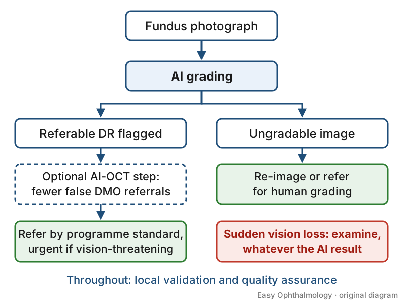
- OCT as a second step to cut false referrals. In a 2026 RCT in Hong Kong, adding AI-OCT to photograph-based screening reduced false-positive DMO referrals from 69.1% to 24.1%, with no DMO missed among non-referred participants [S11].
- Low-resource deployment. Portable fundus cameras and trained general physicians showed good agreement with reference standards in developing countries [S6]. Success depends on infrastructure, local validation and regulatory support [S7].
- Patient data. Do not upload identifiable patient images or data to unapproved tools; follow your local data-protection rules.
Refer when
| Situation | Urgency |
|---|---|
| AI flags referable or vision-threatening DR | Refer by your programme's standard, urgent for vision-threatening disease |
| Image ungradable by AI | Re-image or refer for human grading ****; timing per your local service or specialist advice |
| Symptoms (sudden vision loss) regardless of AI result | Immediate, same-day [G1] |
Pitfalls
- Applying published accuracy to a different camera or population [S1] [S7].
- Ignoring ungradable images [S1] [S2].
- Trusting internally validated models [S10].
- Treating oculomics predictions as diagnostic [S13].
- Vendor involvement: EyeArt specificity varied with vendor involvement [S4].
Evidence notes
| Source | Date | Design |
|---|---|---|
| [S1] | 2026 | Systematic review and meta-analysis of prospective studies |
| [S2] | 2024 | Systematic review and meta-analysis, real-world settings |
| [S3] | 2025 | Meta-analysis, IDx-DR |
| [S4] | 2026 | Meta-analysis, EyeArt |
| [S5] | 2023 | Meta-analysis, smartphone systems |
| [S6] | 2025 | Systematic review and meta-analysis, developing countries |
| [S7] | 2025 | Umbrella review |
| [S8] | 2024 | RCT, 164 participants [S8] |
| [S9] | 2025 | Meta-analysis, 6 studies [S9] |
| [S10] | 2024 | Meta-analysis, 53 studies [S10] |
| [S11] | 2026 | Prospective validation and RCT |
| [S12] | 2023 | Meta-analysis |
| [S13] | 2024 | Systematic review of bias and reporting |
- Diagnostic-accuracy meta-analyses show high heterogeneity [S12].
- Not covered by these sources: generative AI and large language models, cost-effectiveness and medicolegal responsibility.
Related articles
- OCT interpretation: macula and retinal nerve fibre layer
- Diabetic retinopathy screening programmes
- Smartphone and portable fundus imaging
References
See the numbered source list below. Each marker [S#] links to its PubMed and DOI record.
Sources
- [S1] Wang TW, Luo WT, Tu YK, Chou YB, Wu YT Prospective validation of deep-learning algorithms for diabetic retinopathy screening: A systematic review and meta-analysis. Survey of ophthalmology. (2026). PubMed 41344407 · doi:10.1016/j.survophthal.2025.11.012
- [S2] Joseph S, Selvaraj J, Mani I, Kumaragurupari T, Shang X, Mudgil P, et al. Diagnostic Accuracy of Artificial Intelligence-Based Automated Diabetic Retinopathy Screening in Real-World Settings: A Systematic Review and Meta-Analysis. American journal of ophthalmology. (2024). PubMed 38438095 · doi:10.1016/j.ajo.2024.02.012
- [S3] Khan Z, Gaidhane AM, Singh M, Ganesan S, Kaur M, Sharma GC, et al. Diagnostic Accuracy of IDX-DR for Detecting Diabetic Retinopathy: A Systematic Review and Meta-Analysis. American journal of ophthalmology. (2025). PubMed 39986640 · doi:10.1016/j.ajo.2025.02.022
- [S4] Wang TW, Luo WT, Tu YK, Chou YB, Wu YT Diagnostic Accuracy of EyeArt for Fundus-Based Detection of Diabetic Retinopathy: A Systematic Review and Meta-analysis. American journal of ophthalmology. (2026). PubMed 41052568 · doi:10.1016/j.ajo.2025.09.045
- [S5] Hasan SU, Siddiqui MAR Diagnostic accuracy of smartphone-based artificial intelligence systems for detecting diabetic retinopathy: A systematic review and meta-analysis. Diabetes research and clinical practice. (2023). PubMed 37805002 · doi:10.1016/j.diabres.2023.110943
- [S6] Yudistira Y, Hendrawan KA, Andayani A, Suryathi NMA, Ernawati T, Gunawan ACV, et al. Diabetic Retinopathy Screening Approaches in Developing Countries: A Systematic Review and Meta-Analysis. Turkish journal of ophthalmology. (2025). PubMed 41142633 · doi:10.4274/tjo.galenos.2025.32916
- [S7] Kabunga R, Asasira J, Njuki S, Daniel A, Morley K, Morley M, et al. Describing the Performance and the Infrastructure Requirements of the Existing Artificial Intelligence (AI)-Based Diabetic Retinopathy (DR) Screening Algorithms for Diabetic Patients: an Umbrella Review. Journal of medical systems. (2025). PubMed 41165895 · doi:10.1007/s10916-025-02280-2
- [S8] Wolf RM, Channa R, Liu TYA, Zehra A, Bromberger L, Patel D, et al. Autonomous artificial intelligence increases screening and follow-up for diabetic retinopathy in youth: the ACCESS randomized control trial. Nature communications. (2024). PubMed 38212308 · doi:10.1038/s41467-023-44676-z
- [S9] Rahmati M, Smith L, Piyasena MP, Bowen M, Boyer L, Fond G, et al. Artificial Intelligence improves follow-up appointment uptake for diabetic retinal assessment: a systematic review and meta-analysis. Eye (London, England). (2025). PubMed 40447778 · doi:10.1038/s41433-025-03849-4
- [S10] Lam C, Wong YL, Tang Z, Hu X, Nguyen TX, Yang D, et al. Performance of Artificial Intelligence in Detecting Diabetic Macular Edema From Fundus Photography and Optical Coherence Tomography Images: A Systematic Review and Meta-analysis. Diabetes care. (2024). PubMed 38241500 · doi:10.2337/dc23-0993
- [S11] Zhang S, Ran A, Zhou J, Ling A, Sham K, Zhang Y, et al. An AI-Based OCT System to Detect Diabetic Macular Edema: A Prospective Validation and Noninferiority Randomized Clinical Trial. JAMA. (2026). PubMed 42295755 · doi:10.1001/jama.2026.7025
- [S12] Leng X, Shi R, Wu Y, Zhu S, Cai X, Lu X, et al. Deep learning for detection of age-related macular degeneration: A systematic review and meta-analysis of diagnostic test accuracy studies. PloS one. (2023). PubMed 37023082 · doi:10.1371/journal.pone.0284060
- [S13] Li Y, Zhang R, Dong L, Shi X, Zhou W, Wu H, et al. Predicting systemic diseases in fundus images: systematic review of setting, reporting, bias, and models' clinical availability in deep learning studies. Eye (London, England). (2024). PubMed 38238576 · doi:10.1038/s41433-023-02914-0
Guidelines
- [G1] College of Optometrists (Guidance for Professional Practice). Annex 4 Urgency of referrals table (n.d.). https://www.college-optometrists.org/clinical-guidance/guidance/guidance-annexes/annex-4-urgency-of-referrals-table